

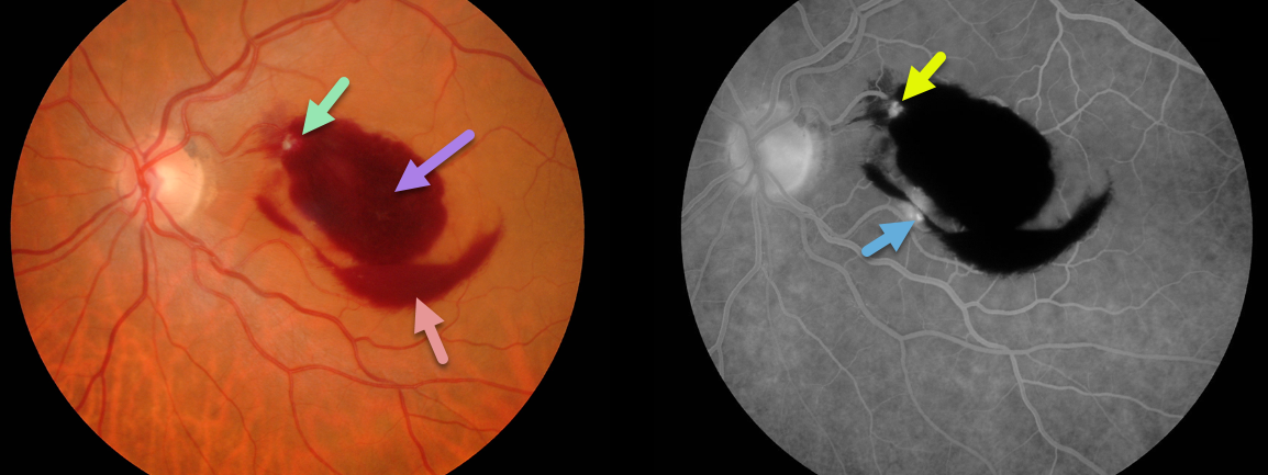
Our patient’s retinal arterial macroaneurysm (RAM) is seen within the superonasal portion of the macular blood. Preretinal and dense intraretinal blood are the clues that a RAM is the cause.
The RAM is confirmed on fluorescein angiography. Although usually singular, our patient may also have a second RAM inferiorly.
Learning Points:
A retinal arterial macroaneurysm (RAM) is thought to occur in a weakened arterial wall from arteriosclerosis.
A RAM appears as a yellow-white dilated lesion along a retinal arterial within the first 3 bifurcations from the optic nerve.
There are 2 distinct presentations, hemorrhagic or exudative. The hemorrhagic type often presents as a pathognomonic multilayer macular hemorrhage, with preretinal, intraretinal and subretinal blood.



