

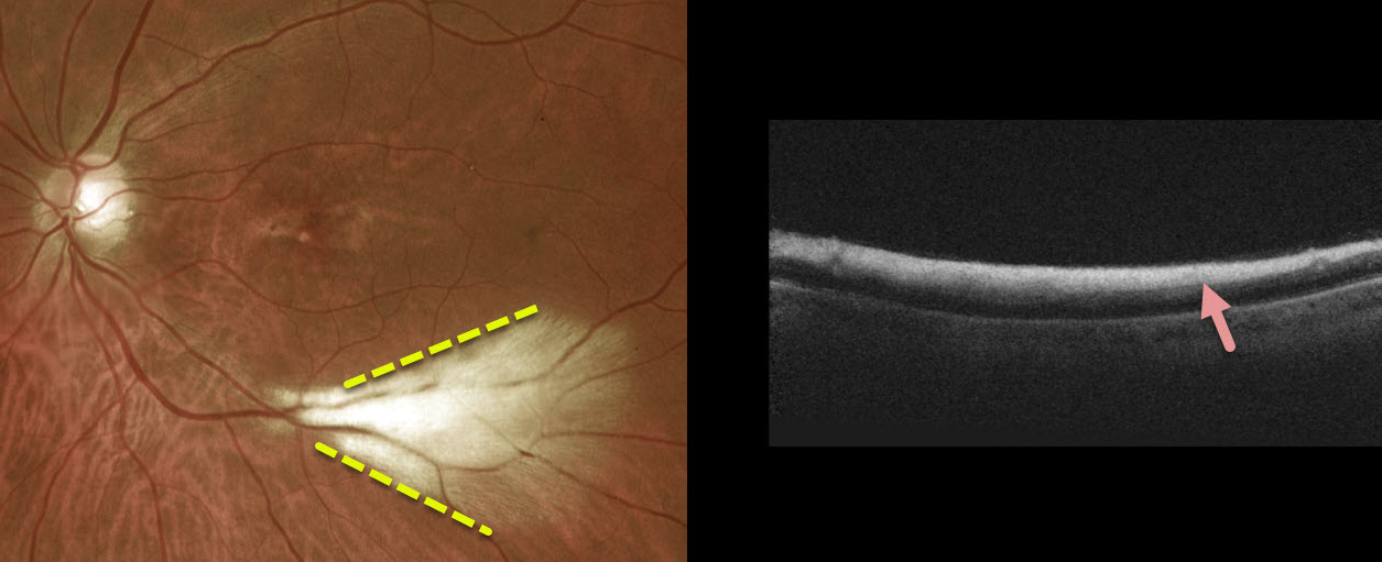
This patient had a large area of myelinated nerve fiber layer (NFL) in the distal inferotemporal left macula. OCT scanning through this area showed marked inner retinal hyperreflectivity.
Learning Points:
Myelinated NFL is a relatively uncommon and usually isolated finding present at birth. Most patients are asymptomatic, although some may have a corresponding relative scotoma.
Myelination can progress in about 10% of cases and can also resolve following retinal ischemic events.
Myelinated NFL occurs when retinal nerve fibers develop a myelin sheath, which usually stops posterior to the lamina cribrosa. The myelination may represent an oligodendrocytic choristoma (Rao et al, Retina 2019;39:1125-1132).



