

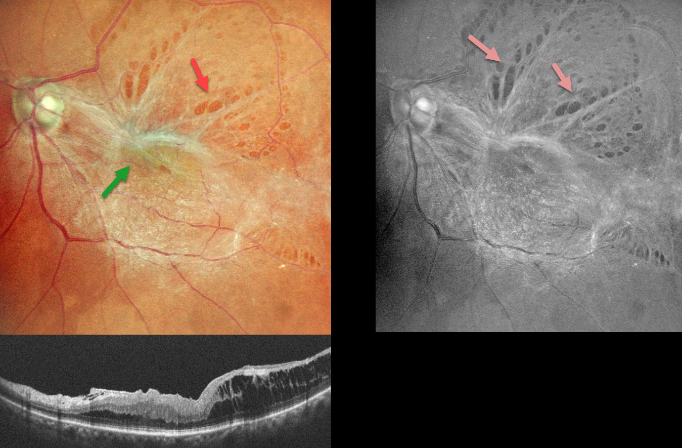
This 58YO female presented with 6 months of vision loss in her left eye. Vision was 20/60 OS.
Color SLO imaging shows a taut epiretinal membrane (ERM) with multiple paravascular lamellar defects, which are better visualized on red-free imaging. OCT scanning shows a stage 3 macular pucker with ectopic inner foveal layer (EIFL) and temporal inner layer schisis.
Learning Points:
An ERM is a thin, fibrous membrane that forms on the retinal surface, often causing central vision loss with metamorphopsia. It can be classified into 4 OCT-based stages (Gonzalez-Saldivar et al, Retina 2020;40:710-717): Stage 1 ERM with preserved foveal contour, Stage 2 Loss of foveal depression with thickened outer nuclear layer, Stage 3 EIFL, and Stage 4 EIFL with complete loss of inner retinal anatomy.
ERMs can cause paravascular lamellar defects due to shearing forces at the vessel borders, and these defects can extend towards the fovea. Our patient’s highly contractile membrane caused multiple striking lamellar defects visualized on fundus photography.



