

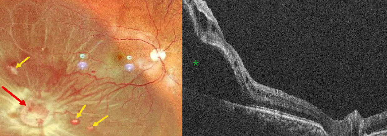
This 52YO male presented with 5 days of vision loss OD. There was a history of bilateral vision loss following facial trauma from a motor vehicle accident 20 years earlier. He denied any family history of eye disease or cancer. Vision was counting fingers OD and 20/200 OS.
Pseudocolor SLO imaging shows an orange-yellow mass consistent with a retinal capillary hemangioma in the inferotemporal midperiphery, causing an exudative retinal detachment. Several smaller hemangiomas are noted. The macula-off detachment is confirmed on OCT scanning.
A recent MRI showed bilateral optic atrophy, a partially empty sella, and an old, healed fracture of the right lateral wall of the orbit. Although emergent vitrectomy surgery was recommended, he was immediately lost to follow-up.
Learning Points:
Retinal capillary hemangiomas are orange-red lesions associated with a prominent paired feeding and draining vessel. These benign vascular tumors need to be aggressively treated since they can cause severe vision loss or blindness from exudative retinal detachment, as in our patient. The presence of hydration outer retinal folds is one of the key findings that help differentiate rhegmatogenous from exudative and traction retinal detachments (Oquendo et al, AJO 2024;268:212-221). The absence of these folds in our patient is typical of an exudative detachment. Common treatment options include thermal laser, cryotherapy, and anti-VEGF injections.
Retinal capillary hemangioma is a predominant feature of VHL, an autosomal dominant condition caused by mutations in the VHL tumor suppressor gene. Patients develop benign and malignant tumors, including central nervous system hemangioblastoma, pheochromocytoma, and renal cell carcinoma.



