

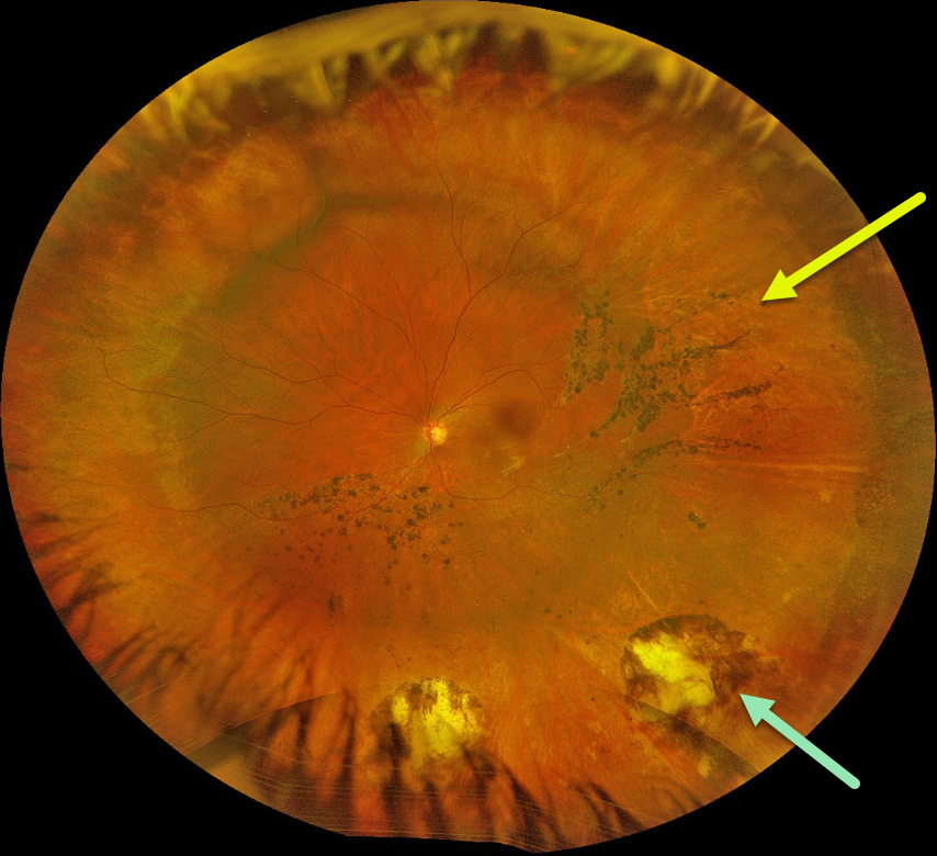
This 31YO female underwent successful scleral buckling surgery about 7 years earlier. Optos color RGB imaging shows a broad encircling buckle. Cryotherapy scarring is noted inferior to the buckle. Multiple demarcation lines with some intraretinal pigment migration are noted within the region of the prior detachment.
Learning Points:
The normal neurosensory retina remains attached to the RPE due to numerous physiologic mechanisms, including the inner (retinal vascular endothelium) and outer (tight junctions at the apex of the RPE cells) blood retinal barriers, hyperosmotic choroid passively drawing fluid out of the subretinal space, RPE pump, interphotoreceptor matrix, and friction between the RPE microvilli and the photoreceptor outer segments. Rhegmatogenous retinal detachment (RRD) occurs when liquid vitreous, entering the subretinal space through a retinal break, overwhelms these forces, favoring retinal detachment.
Repairing RRD involves finding and closing all breaks, essentially denying the liquid vitreous access to the subretinal space. The break(s) can be closed internally with a gas bubble with pneumatic retinopexy or vitrectomy surgery, or externally by sewing a piece of silicone material to the eye wall (scleral buckle). This indents, or buckles, the RPE and sclera up against the retinal break and formed vitreous.
The choice of procedure to reattach the retina is complex, depending on each patient’s unique anatomy and the surgeon’s preference. Historically, scleral buckling was the only method until vitrectomy surgery appeared in the 1970’s. There are advantages and disadvantages to all techniques. With one or more surgeries, there is a high anatomic success rate regardless of the method of retinal reattachment.



