

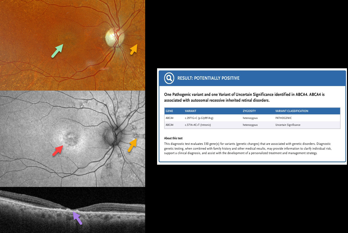
This 88YO female was diagnosed with age-related macular degeneration elsewhere. Vision was 20/400 OD and 20/50 OS.
Optos color RGB imaging OD shows a round area of subfoveal pigmentary changes with extensive drusen-like lesions extending outwards from the peripheral macula. Fundus autofluorescence (FAF) shows central hypo-FAF with a ring of hyper-FAF around this foveal lesion. Scattered lesions consisting of small dots of hyper-FAF with surrounding hypo-FAF are scattered around the nerve. Swept-source OCT shows severe central neurosensory atrophy with a macular pseudohole. Some clumps of hyperreflective material are noted within the area of atrophy, along with variable ellipsoid zone loss elsewhere. Similar findings were present in her left eye (not shown). Genetic testing revealed heterozygous ABCA4 mutations, including a pathogenic variant (c.2971G>C, p.Gly991Arg) and a variant of uncertain significance (c.5714.+4C-T, intronic).
Learning Points:
The ABCA4 protein is located in the photoreceptor outer segments and is involved in the recycling of 11-cis-retinal. The mutation of this gene causes the accumulation of lipofuscin in the RPE, which eventually leads to photoreceptor and RPE degeneration. ABCA4 disorders include Stargardt disease, fundus flavimaculatus, cone-rod dystrophy, retinitis pigmentosa, and age-related macular degeneration.



