

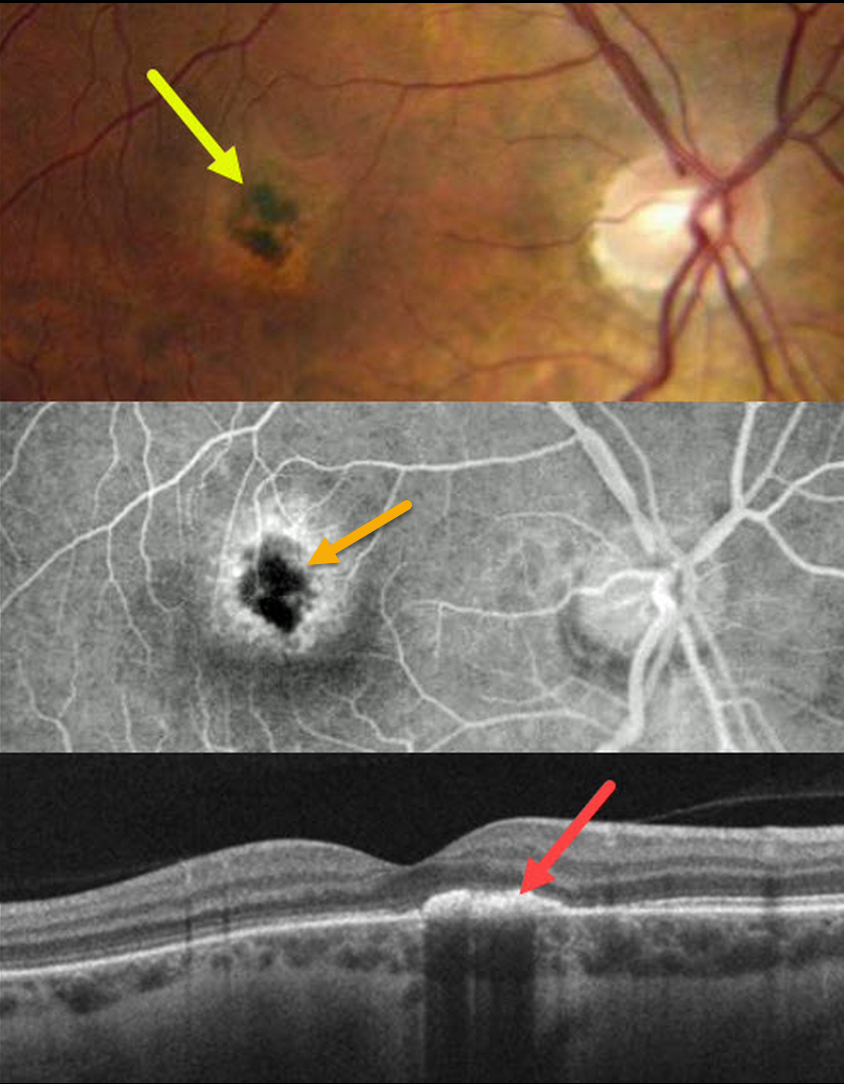
This 33YO healthy female complained of 1 to 2 weeks of a blind spot and ring of light in her right eye. She had a recent non-COVID viral illness when her symptoms started. Vision was 20/60 OD and 20/20 OS.
Color imaging shows a subfoveal area of central hyperpigmentation surrounded by a ring of pigment loss. Angiographically, the pigmentation blocks with a surrounding rim of staining.
OCT B-scan shows a hyperreflective plaque in the outer retina.
Learning Points:
Acute idiopathic maculopathy (AIM) is an idiopathic inflammation of the outer retina, RPE, and inner choroid. There is often a preceding viral prodrome caused by the Coxsackie virus. Healthy young patients typically present with acute unilateral moderately severe central vision loss.
The initial funduscopic changes show macular fluid and, sometimes, deep retinal blood, which can simulate macular neovascularization. The OCT shows heterogeneous hyperreflective thickening of the outer retina/RPE and fluid.
Fluorescein angiography usually shows deep retinal leakage. The lesion spontaneously resolves with significant visual improvement after several weeks, usually leaving a pathognomonic bull’s-eye lesion as seen in our patient.



