

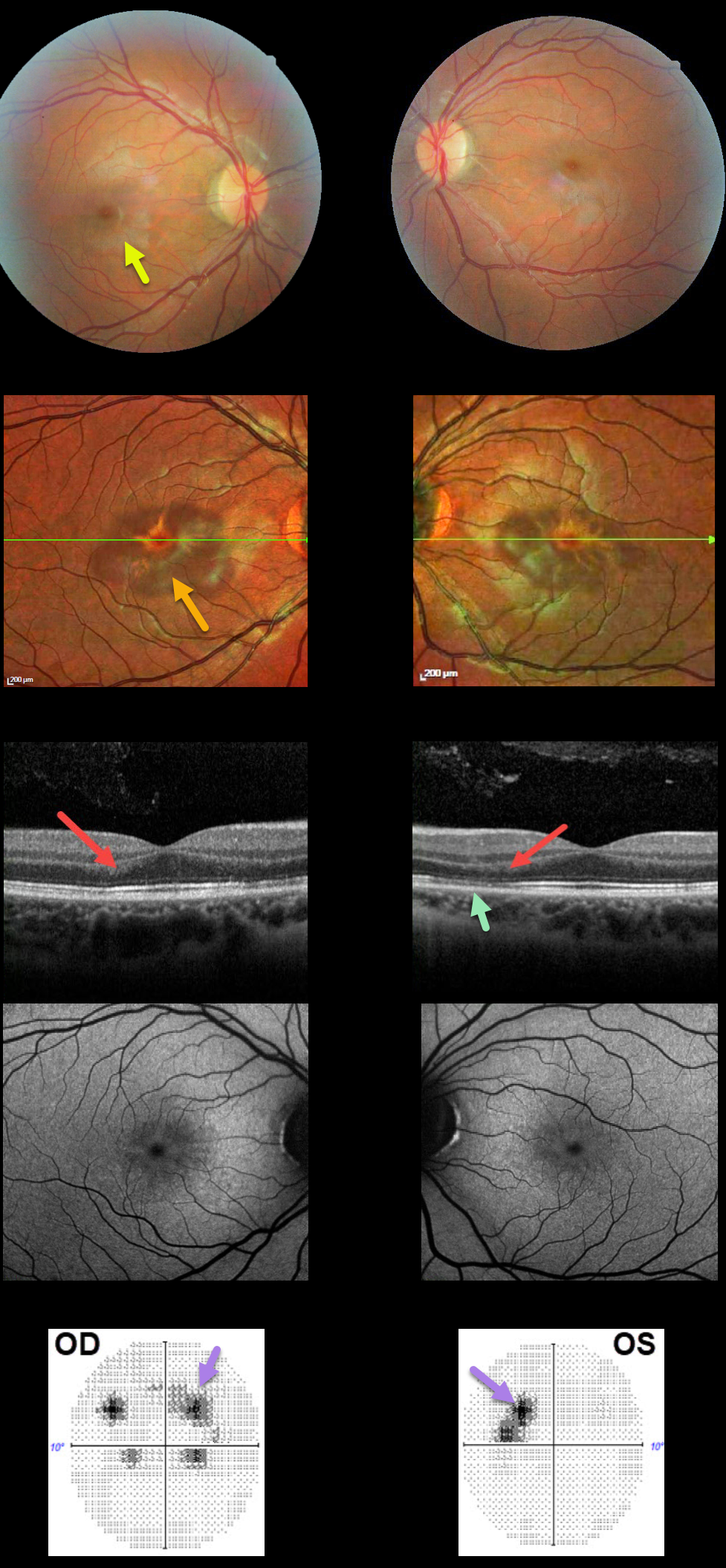
This 21YO female presented with a two-week history of bilateral paracentral relative scotomas. This was preceded by myalgias, sinus pressure, sore throat, and a cough. There was a 4-year history of oral contraceptive use. Vision was 20/25 OU.
There are faint reddish-brown wedge-shaped lesions radiating out of each fovea, seen more easily on multicolor imaging.
OCT scanning shows some focal areas of increased hyperreflectivity in the outer nuclear layer and defects in the ellipsoid zone. Fundus autofluorescence is relatively normal.
10-2 visual fields confirm the paracentral scotomas noticed by the patient. Over the following months, her 10-2 visual field partially improved, coinciding with partial normalization of the OCT findings (not shown).
Learning Points:
This case highlights many characteristic features of acute macular neuroretinopathy (AMN), an idiopathic disorder likely caused by compromise of the deep retinal capillary plexus.
Multicolor and infrared imaging often highlight the typical wedge-shaped AMN lesions more effectively than color fundus photos.
84% of patients are female with a median age of 30 years, and 80% are non-Latino white.
The most common association is a flu-like prodrome (48%), followed by oral contraceptive use (36%).
Presenting acuity is 20/40 or better in 80% of eyes, coinciding with sparing of the sub-foveal areas. Roughly half of AMN cases are bilateral. There is typically moderate visual improvement over the coming months, and no treatment is recommended.
For a great summary of AMN, see Bhavsar KV et al, Surv Ophthalmology 2016; 61:538-565.



