

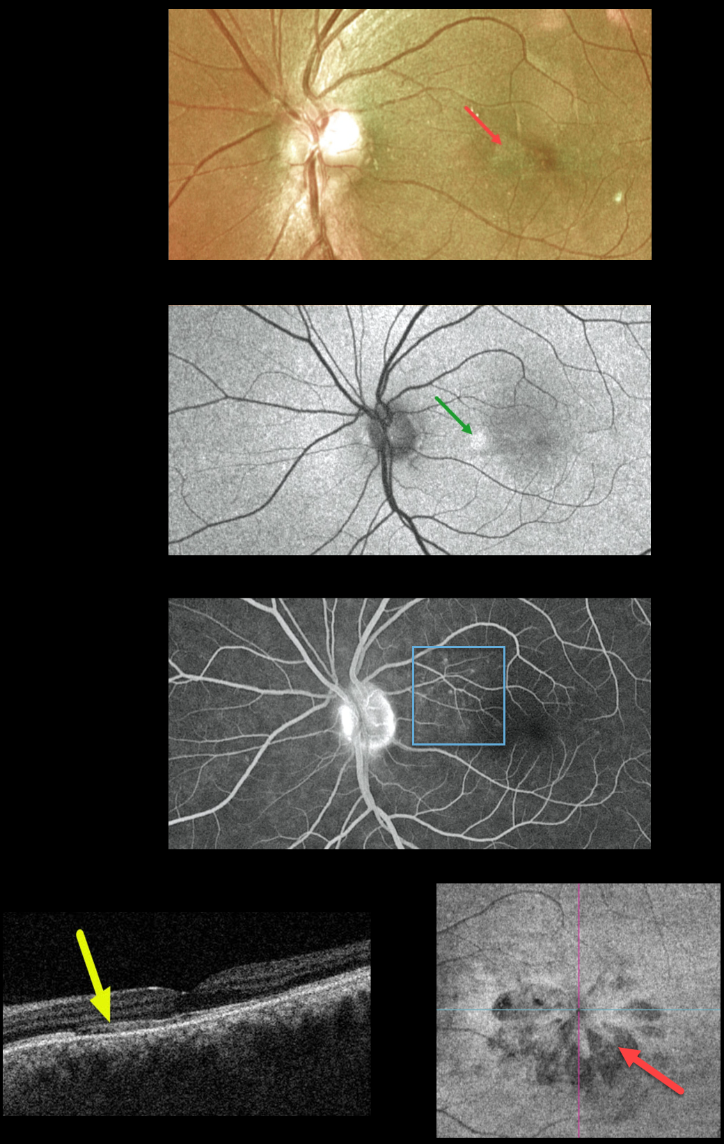
This 39YO male presented with 1 day of blurred vision in his left eye. Vision was 20/30 OU. Optos color imaging shows a wedge-shaped lesion radiating into the nasal fovea. Optos fundus autofluorescence (FAF) shows hyper-FAF in this area, and fluorescein angiography shows subtle areas of increased nasal macular hyperfluorescence.
Swept-source OCT shows attenuation of the ellipsoid zone and outer segment layers. En face imaging of the outer retina shows multiple hyporeflective wedge-shaped lesions radiating from the macular center. Findings in the right eye were completely normal.
Learning Points:
This case highlights many characteristic features of acute macular neuroretinopathy (AMN), an idiopathic disorder likely caused by compromise of the deep retinal capillary plexus.
Multicolor and infrared imaging often highlight the typical wedge-shaped AMN lesions more effectively than color fundus photos, although the Triton en face OCT image in our patient beautifully imaged these.
Most patients are white, non-Latino females with a median age of 30 years. Roughly half of AMN cases are bilateral. The most common associations are a flu-like prodrome and oral contraceptive use.
Presenting acuity is usually good, coinciding with sparing of the sub-foveal areas. There is typically moderate visual improvement over several months without treatment.
For a great AMN review, see Bhavsar et al, Surv Ophthalmology 2016;61:538-565.



