

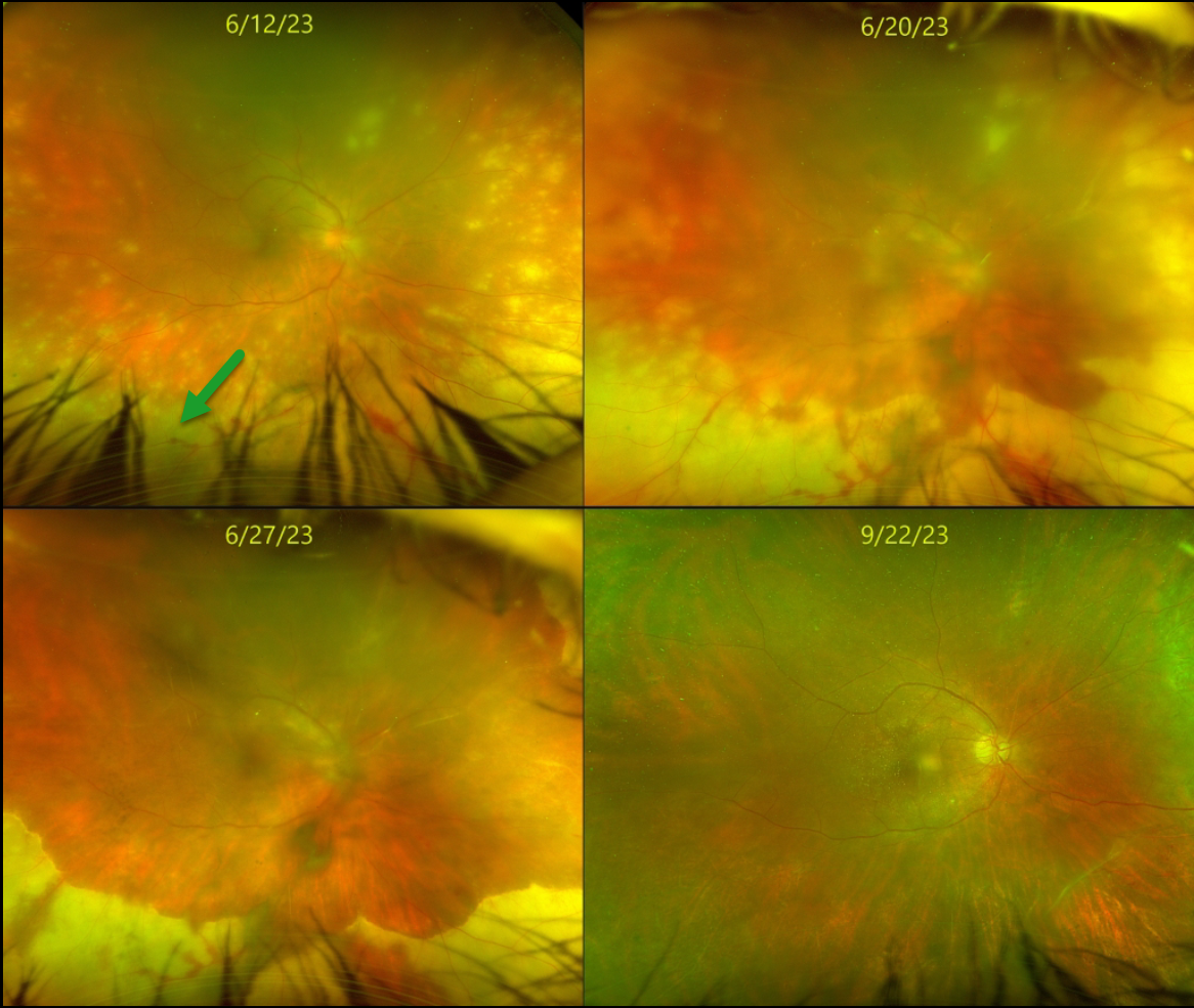
This healthy 55YO female presented with about 2 weeks of vision loss in her right eye. Vision was counting fingers OD and 20/30 in her normal left eye.
Slit lamp showed moderate anterior granulomatous inflammation. Moderate vitreous debris was noted. Optos color RG imaging shows multifocal white patches of variably confluent outer retinal inflammation extending anteriorly from the midperiphery. She was started on topical steroids and atropine, valacyclovir 1 gram PO BID, and a baby aspirin daily for acute retinal necrosis syndrome (ARNS).
After 1 week, vision remained at counting fingers but with subjective improvement. The retinitis was consolidating. The retinitis gradually improved over the following 3 months. When last examined on 9/22/23, vision was 20/70, with complete resolution of disease on valacyclovir 1 gram PO daily. We continue to follow her closely.
Learning Points:
ARNS is most commonly caused by the Herpes zoster virus. It usually occurs in immunocompetent individuals, and is a rapidly progressive panuveitis with hemorrhagic and ischemic unilateral (ARNS) or bilateral (BARNS) retinal necrosis that typically begins in the retinal periphery.
ARNS is often a devastating and blinding infection due to the high risk for retinal detachment as well as macular and optic nerve involvement. The latest meta-analysis argues for systemic antiviral therapy and prophylactic vitrectomy, with uncertain benefits for prophylactic laser photocoagulation or adjunctive intravitreal antivirals (see Zhao et al, Retina 2021;41:965-978).



