

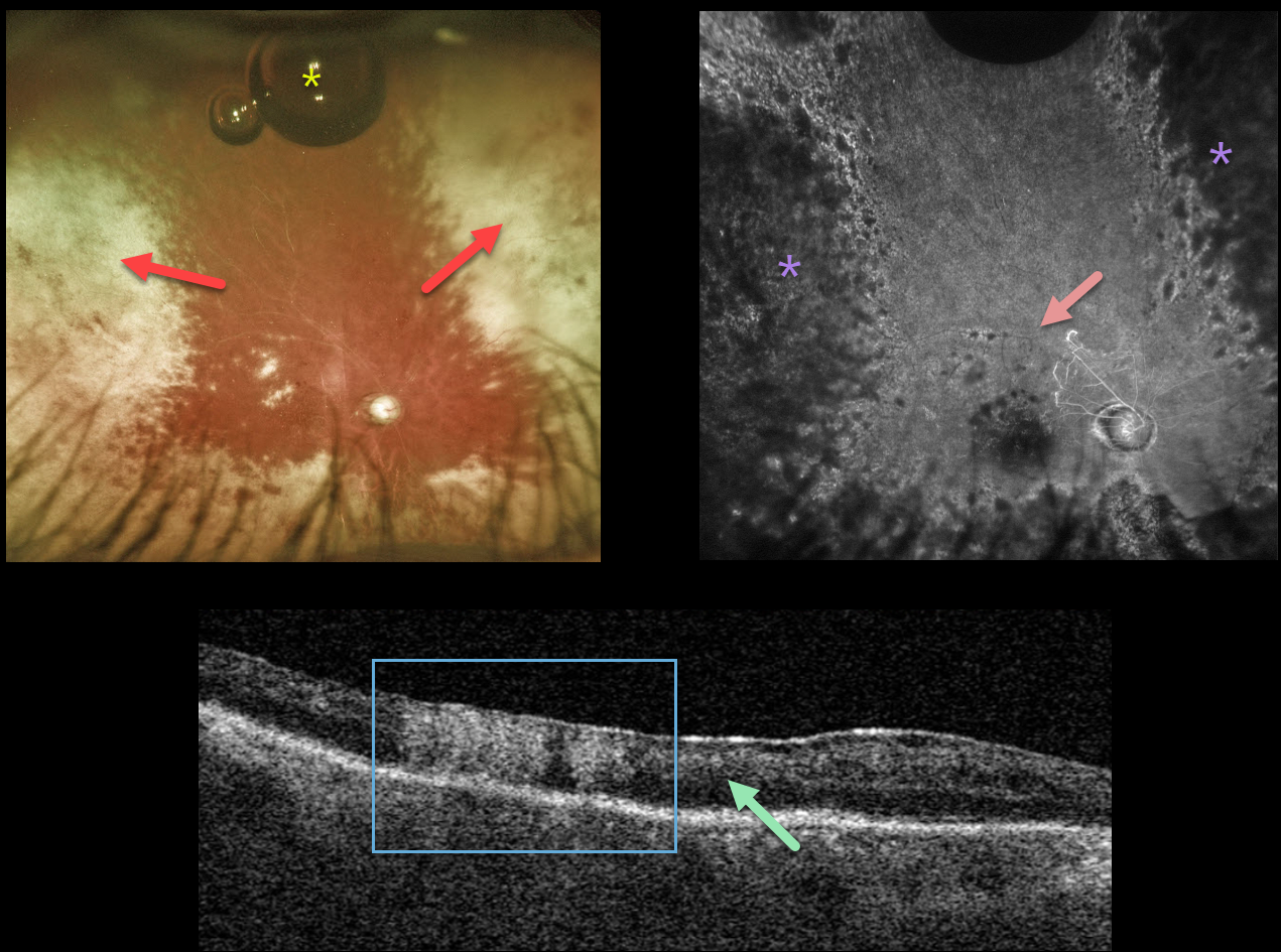
This 58YO male has an extremely complicated past ocular history beginning with pseudoexfoliative glaucoma with a steroid response. Following cataract surgery in 2012, his intraocular lens dislocated in February 2016, requiring vitrectomy with a secondary anterior chamber implant. Pseudophakic bullous keratopathy developed, requiring DMEK surgery in July 2018.
His intraocular pressure subsequently increased, eventually requiring multiple glaucoma procedures, including Baerveldt implantation. Additional DMEK procedures were performed in September 2020 and December 2021. A minor additional corneal procedure with injection of gas into the anterior chamber was performed on 5/5/22 for some host Descemet’s that extended under the donor graft, causing recurrent corneal edema.
When examined on 6/24/22, the cornea was finally clear enough to allow for visualization of the posterior segment, which hadn’t been possible during his multiple anterior segment procedures. Vision was light perception.
Optos imaging shows white chorioretinal scarring in the nasal and temporal peripheries with a small amount of residual intraocular gas that had migrated into the vitreous cavity. Triton swept source OCT shows diffuse disorganization of all retinal layers, with some areas of full-thickness hyperreflectivity. Fluorescein angiography shows near total loss of all retinal perfusion, with peripheral hypofluorescence from the white peripheral chorioretinal scarring.
We felt his findings were most consistent with acute retinal necrosis, and oral valaciclovir was started.
Learning Points:
ARNS is most commonly caused by the Herpes zoster virus. It usually occurs in immunocompetent individuals, and is a rapidly progressive panuveitis with hemorrhagic and ischemic unilateral (ARNS) or bilateral (BARNS) retinal necrosis that typically begins in the retinal periphery.
ARNS is often a devastating and blinding infection due to the high risk for retinal detachment as well as macular and optic nerve involvement. The latest meta-analysis argues for systemic antiviral therapy and prophylactic vitrectomy, with uncertain benefits for prophylactic laser photocoagulation or adjunctive intravitreal antivirals (see Zhao et al, Retina 2021;41:965-978).



