

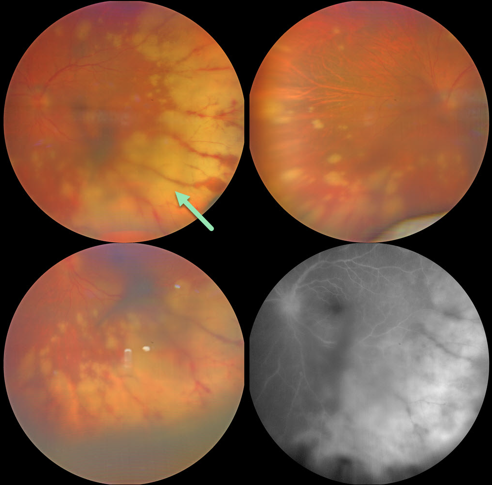
This 56YO female presented with 2 weeks of decreased vision in her left eye. She was treated for herpes zoster one month earlier, and there were a few crusted lesions on the right side of the forehead. Vision was 20/20 in her normal OD and 20/200 OS. There were moderate anterior chamber and vitreous cells.
Color photography shows scattered, midperipheral, variably confluent patches of creamy white deep retinitis. Fluorescein angiography shows optic nerve and deep retinal leakage.
An anterior chamber tap was performed, and PCR analysis confirmed varicella zoster. Intravenous acyclovir was given for 1 week, followed by oral valacyclovir 1 gram daily. When examined 3 weeks later, vision improved to 20/40 with resolving retinitis (not shown).
Learning Points:
The acute retinal necrosis syndrome is usually caused by the herpes zoster virus. It generally occurs in immunocompetent individuals and presents as a rapidly progressive panuveitis with hemorrhagic and ischemic unilateral (ARNS) or bilateral (BARNS) retinal necrosis, typically beginning in the retinal periphery.
It is often a devastating and blinding infection due to the high risk for retinal detachment as well as macular and optic nerve involvement. The latest meta-analysis argues for systemic antiviral therapy and prophylactic vitrectomy, with uncertain benefits for prophylactic laser photocoagulation or adjunctive intravitreal antivirals (Zhao et al, Retina 2021;41:965-978).
Treatment should be initiated as soon as the diagnosis is suspected, without waiting for laboratory results. Oral valacyclovir is clinically equivalent to intravenous acyclovir (Baltinas et al, AJO 2018;188:173-180), and aspirin is often recommended to minimize vascular thrombosis. Treatment is continued until all retinitis becomes inactive, and usually for at least 3-6 months thereafter to reduce the risk of fellow-eye involvement.



