

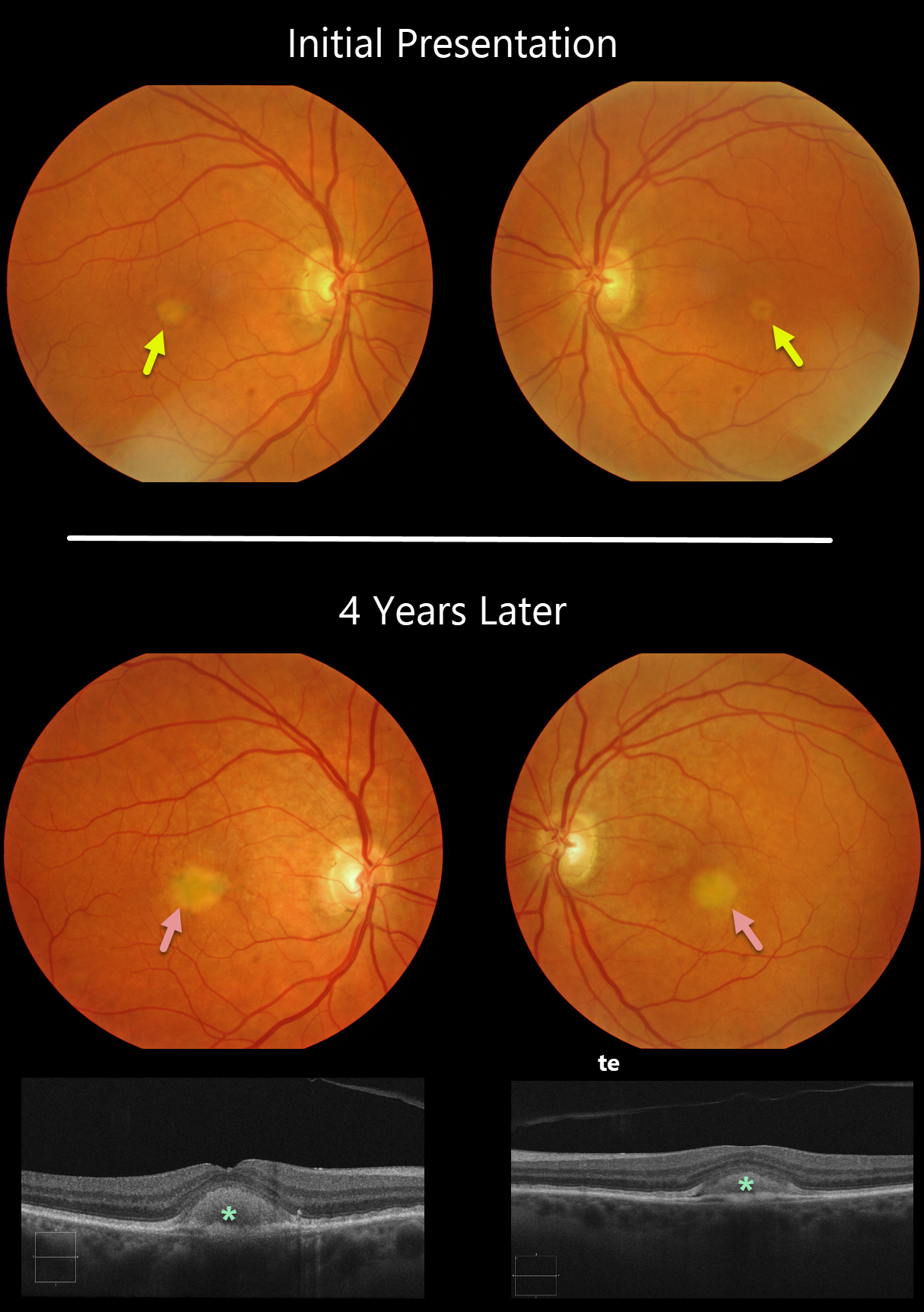
Our patient shows classic findings for adult-onset foveomacular vitelliform dystrophy (Adult Best’s), including small, round, symmetrical subfoveal yellow spots. The vitelliform lesions enlarged over four years, which is seen on OCT as subretinal hyperreflective material between the RPE and outer retina. This material often indents the overlying retina.
Learning Points:
Adult Best’s is associated with a mutation in the BEST1 gene, which encodes the bestrophin-1 protein. Bestrophin-1, a calcium-activated chloride channel, is primarily found in the basolateral plasma membrane of the RPE.
BEST1 mutations cause a variety of phenotypes, including autosomal recessive bestrophinopathy, best vitelliform macular dystrophy, and autosomal dominant vitreoretinochoroidopathy.
These patients tend to maintain fairly good vision. Outer retinal and RPE atrophy can cause central vision loss, as can the rare development of macular neovascularization. Patients should therefore regularly monitor their vision with an Amsler grid.



