

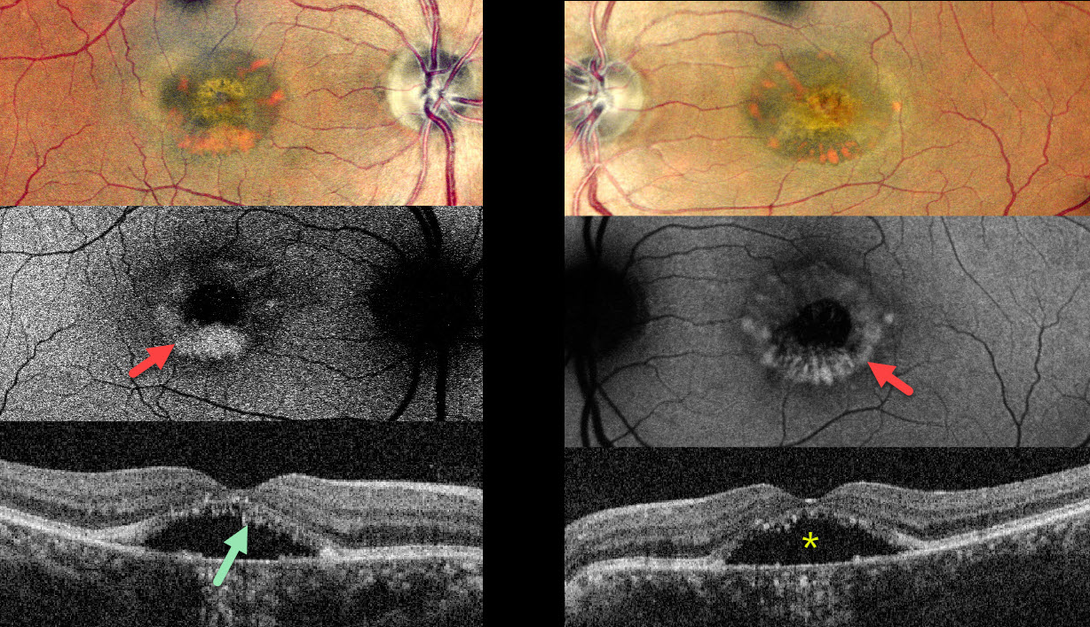
This 62YO female complained of stable bilateral blurred vision for seven years, having been diagnosed with central serous chorioretinopathy elsewhere. Vision was 20/30 OD and 20/40 OS.
Pseudocolor SLO imaging shows symmetrical, partially scrambled vitelliform lesions. The lesions are hyperautofluorescent along their outer edges and hypoautofluorescent centrally. OCT shows shaggy hyperreflective material lining the outer retina overlying foveal subretinal fluid.
Learning Points:
Adult-onset foveomacular vitelliform dystrophy is characterized by bilateral, symmetrical, grayish-yellow, one-third to one-half disc diameter subfoveal lesions with a central pigmented spot. In our experience, it is the most common pattern of dystrophy and closely resembles Best disease, although the lesions are generally smaller and diagnosed in adulthood. Causative mutations include BEST1, PRPH2, IMPG1 and IMPG2.
For a great review article, see Chowers et al, Progress in Retinal and Eye Research 2015;47:64-85.



