

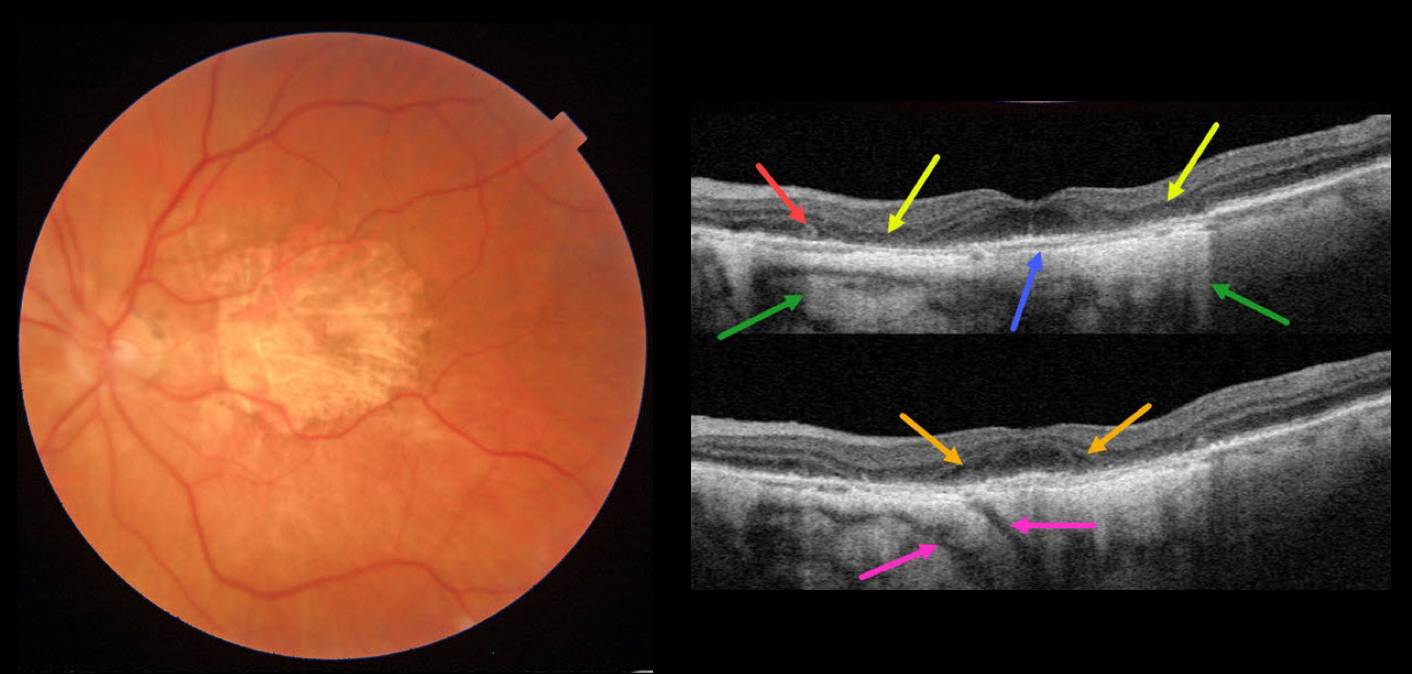
This 84YO female has stable advanced non-exudative AMD with bilateral macular atrophy (MA). There are numerous classic findings on multimodal imaging of her left eye.
Color imaging shows foveal MA. Swept-source OCT shows pericentral outer retinal and RPE atrophy. This atrophy allows for more light to illuminate the underlying choroid and sclera, as well as visualizing two short posterior ciliary arteries posterior to the sclera. The variably atrophic RPE allows for visualization of Bruch membrane, which is normally fused in the RPE-Bruch layer seen in normal eyes. There is a tiny area of outer retinal tubulation (ORT) nasally. Finally, hyporeflective outer plexiform layer wedge defects are noted along the descending outer retinal atrophy.
Wedge defects develop at the boundaries of the degenerating outer plexiform layer in about 75% of MA eyes (Mones et al, Ophthalmology 2012;119:1412-1419).
ORTs represent photoreceptor and Muller cell degeneration (Dolz-Marco et al, Ophthalmology 2017;124:1353-1367). These ORTs have a hyper-reflective border with a central hyporeflective core.
Learning Points:
In 1970, Dr. Gass originally described macular atrophy from AMD as “geographic areas of atrophy” in the setting of “senile macular choroidal degeneration” (Schmitz-Valkenberg, Retina 2016;36:2250-2264).
Today, the terminology is much more specific due to advances in technology and improved visualization of retinal and choroidal structures.
From Sadda et al, Ophthalmology 2018:537-548, the new classification is as follows: Complete RPE and Outer Retinal Atrophy (cRORA), Incomplete RPE and Outer Retinal Atrophy (iRORA), Complete Outer Retinal Atrophy (cORA), and Incomplete Outer Retinal Atrophy (iORA).



