

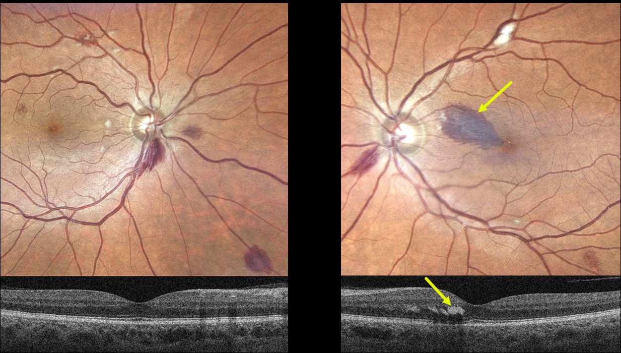
This 55YO male presented with a one-week history of a paracentral scotoma in his left eye. Vision was 20/40 OD and 20/60 OS. He gave a history of aplastic anemia, and his hemoglobin was 6.2 when he developed these symptoms. Blood transfusion brought his hemoglobin to over 8. He told us that his blood pressure runs low to normal.
Optos imaging shows scattered bilateral deep and superficial retinal hemorrhages with a few cotton wool spots. A tear-drop-shaped, larger hemorrhage is noted in the superonasal left macula. This hemorrhage localizes to the outer plexiform layer on swept-source OCT.
Learning Points:
The central retinal artery supplies the inner two-thirds of the retina, while the choroid supplies the outer third (the photoreceptor layers), the RPE, and the choroid. The outer plexiform layer (OPL) is the ‘end of the line’ for the inner retinal circulation.
In severe anemia, the more proximal inner retina removes whatever oxygen is available, leaving the deeper layers relatively ischemic. This can cause dysfunction of the retinal vascular endothelial tight junctions, which allows red blood cells to enter and stain the OPL, appearing clinically as deep retinal blot hemorrhages.
Deep blot OPL hemorrhages also appear for similar reasons in the temporal retinal periphery in ocular ischemia.
We advised observation, and expect his findings and symptoms to improve as long as his anemia is controlled.



