

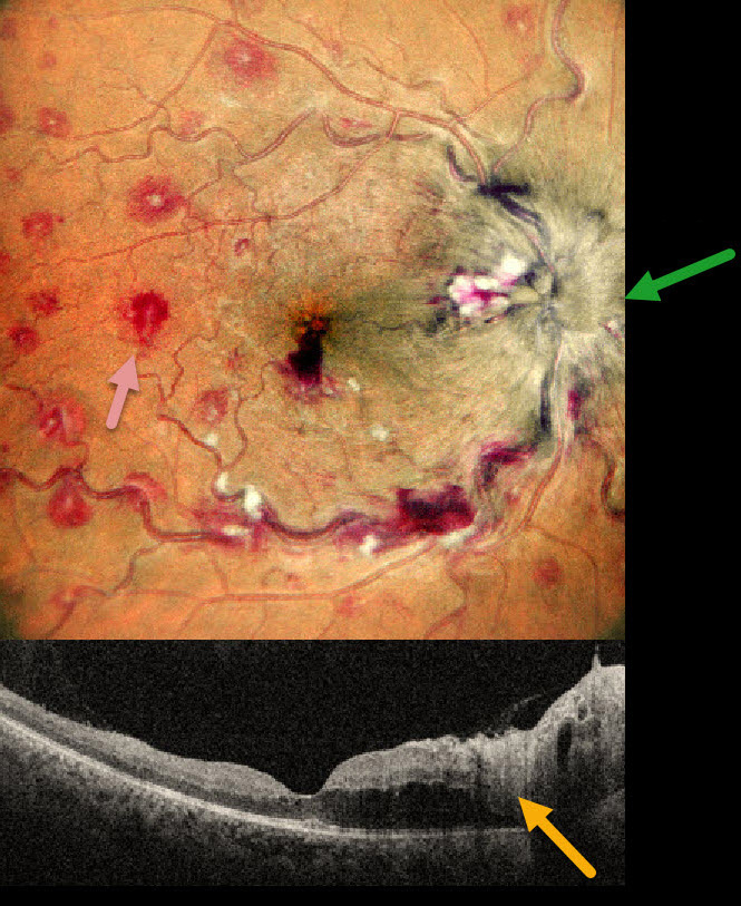
This 35YO male presented with 1 month of vision loss in his right eye. There was no past medical history. Vision was 20/40 OD and 20/20 in his normal OS.
MultiColor imaging shows a markedly swollen optic nerve with extensive multifocal deep, white-centered retinal hemorrhages. OCT shows marked mostly outer retinal edema extending from the nerve towards the macular center.
Extensive laboratory evaluation was negative except for a microcytic anemia with a hemoglobin of 2.9 g/dl. This was felt to be caused by an iron deficiency due to poor diet. Intravitreal anti-VEGF injections were recommended, along with oral prednisone for the optic nerve swelling. Unfortunately, he was immediately lost to follow-up.
Learning Points:
Anemia-related retinal findings most commonly include hemorrhages in the retina and sub-internal limiting membrane. Severe anemia is a rare cause for papilledema and should always be considered in patients with atypical idiopathic intracranial hypertension (Biousse et al, AJO 2003;135:437-446). The underlying pathogenesis of anemia-induced optic nerve swelling remains unknown. It is unclear why our patient had unilateral findings from a systemic disorder.



