

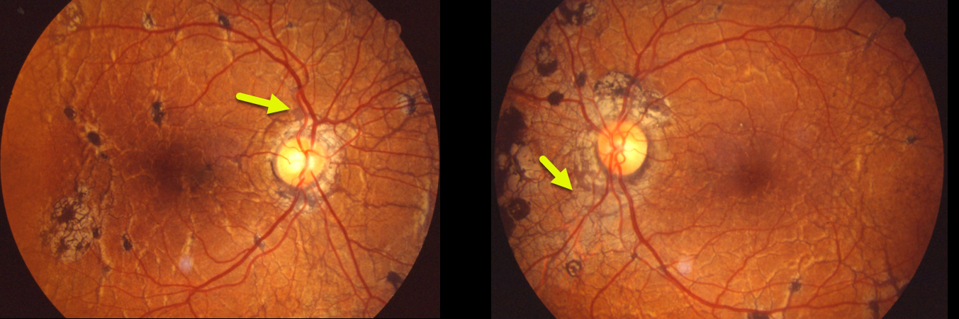
This patient was examined in 1989 and had classic findings of pseudoxanthoma elasticum. Extensive angioid streaks encircle and radiate from the nerves, extending into the peripheral maculas and beyond the arcades.
Angioid streaks are usually less dramatic and are often quite easy to miss since their color and orientation mimic the normal retinal vasculature, hence the term “angioid”.
The orange skin (peaux d’orange) appearance, most pronounced in each temporal macula, is caused by the calcified Bruch’s membrane giving a yellow mottled ‘coquille d’oeuf’ (eggshell) appearance that contrasts with the normal orange RPE and choroidal pigmentation (Spaide, Retina 2015;35:392-397).
Learning Points:
Angioid streaks are known to be associated with systemic conditions such as pseudoxanthoma elasticum, Ehlers-Danlos syndrome, Paget’s disease, sickle cells, or idiopathic (PEPSI).
These patients are at significant risk for macular neovascularization and need to be followed and treated as needed with anti-VEGF agents, as in patients with neovascular AMD.
Genetic testing with pseudoxanthoma would test positive for a pathogenic ABCC6 mutation.



