

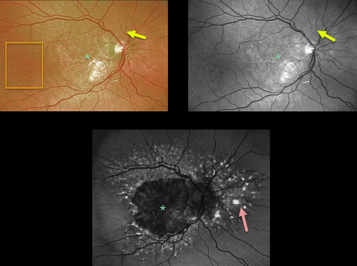
This patient has angioid streaks that radiate from the optic nerve. These are very easy to miss unless they are explicitly looked for.
Optos color and green-channel photographs of the right eye show central macular atrophy with more temporal peaux d’orange changes. The left eye (not pictured) showed identical findings.
Fundus autofluorescence (FAF) shows widespread hyper-FAF lesions surrounding the central hypo-FAF macula. These multifocal hyper-FAF lesions are typical of pattern dystrophies and can somewhat simulate fundus flavimaculatus if they extend outside the macula.
This patient is monitored regularly due to the risk of developing macular neovascularization, although that is fairly unlikely given the macular atrophy.
Learning Points:
Patients with angioid streaks and pseudoxanthoma elasticum (PXE) can present with retinal findings similar to those of pattern dystrophies.
These patients are at significant risk for macular neovascularization (MNV) and need to be followed and treated as needed with anti-VEGF agents, as in patients with AMD.



