

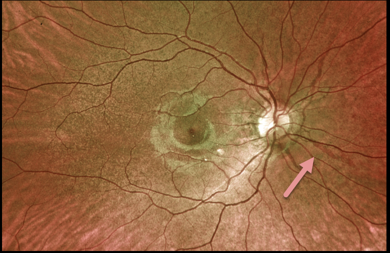
This 19YO female presented with these asymptomatic fundoscopic changes. Vision was 20/25 OU. Optos imaging shows angioid streaks radiating from each optic nerve.
Our patient denied any past medical or family history of pseudoxanthoma elasticum. Genetic testing for the ABCC6 mutation is pending.
Learning Points:
Angioid streaks are often quite easy to miss since their color and orientation mimic the normal retinal vasculature, hence the term ‘angioid.’
The orange skin (peaux d’orange) appearance, most pronounced in each temporal macula, is caused by the calcified Bruch’s membrane giving a yellow mottled ‘coquille d’oeuf’ (eggshell) appearance that contrasts with the normal orange RPE and choroidal pigmentation (Spaide, Retina 2015;35;392-397).
These patients are at significant risk for macular neovascularization (MNV) and need to be followed and treated as needed with anti-VEGF agents, as in patients with AMD.



