

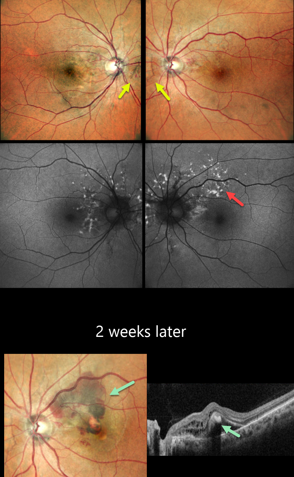
This 45YO male presented for a second opinion for bilateral wavy vision for 2 months. He was diagnosed elsewhere with macular neovascularization (MNV) and received two prior anti-VEGF injections (last 10 days earlier). Vision was 20/20 OU.
Pseudocolor SLO imaging shows subtle bilateral peripapillary angioid streaks associated with a few retinal hemorrhages. Fundus autofluorescence (FAF) shows numerous mostly peripapillary hyper-FAF flecks. Initial observation was recommended.
He returned two weeks later with new symptoms OS, and vision decreased to 20/120. New submacular fluid and blood are noted clinically and on OCT, and monthly anti-VEGF therapy was restarted. Unfortunately, he was lost to follow-up after 3 injections.
Learning Points:
Angioid streaks are orange-yellow, irregular, crack-like dehiscences in Bruch’s membrane associated with atrophic degeneration of the overlying RPE. These patients, especially those with pseudoxanthoma elasticum (PXE), are at significant risk for MNV as occurred in each of our patient’s eyes. MNV responds best to anti-VEGF therapy, and treatment is usually ongoing as with the typical neovascular AMD protocols. However, long-term vision is often poor due to RPE and choroidal atrophy (Mori et al, Graefe’s 2020;258:1863-1869).
Our patient had no family history of PXE or suggestive skin lesions, so skin biopsy or genetic testing was not performed. However, the funduscopic changes and macular dystrophy-like FAF findings are fairly pathognomonic for PXE.



