

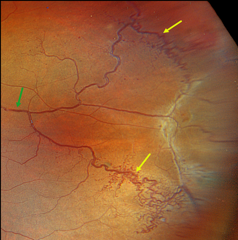
This healthy 5YO boy was referred for an asymptomatic retinal lesion in his left eye. Vision was 20/25 OU.
Optos color RGB imaging shows unusual temporal changes involving the venous vascular tree. Prominent dilated and tortuous retinal veins drain the supero- and inferotemporal periphery before merging into a single vessel more posteriorly.
Surrounding these larger veins, especially inferiorly, are networks of small, corkscrew venules, along with more scattered tiny red microaneurysmal-like structures. Temporally, within the area drained by the larger dilated veins, there is a broad area of variable preretinal fibrosis.
Learning Points:
Retinal cavernous hemangiomas are usually unilateral, inner retinal grape-like clusters of aneurysms filled with dark venous blood. A gray fibrotic membrane often covers its superficial surface.
Fluorescein angiography shows plasma-erythrocyte layering within these saccular lesions caused by sluggish blood flow. Vision is usually normal, and no treatment is required. Rarely can vision loss occur from vitreous hemorrhage.
Although most occur sporadically, some may be associated with an autosomal dominant neurocutaneous syndrome, including cutaneous and central nervous system hemangiomas. Neurologic imaging is therefore recommended.
We believe our patient most likely has an atypical retinal cavernous hemangioma, given the scattered microaneurysmal-like changes (likely small ‘grapes’) and preretinal fibrotic component. Fluorescein angiography would help establish this diagnosis, but this was not possible given our patient’s age. Genetic testing was negative.



