

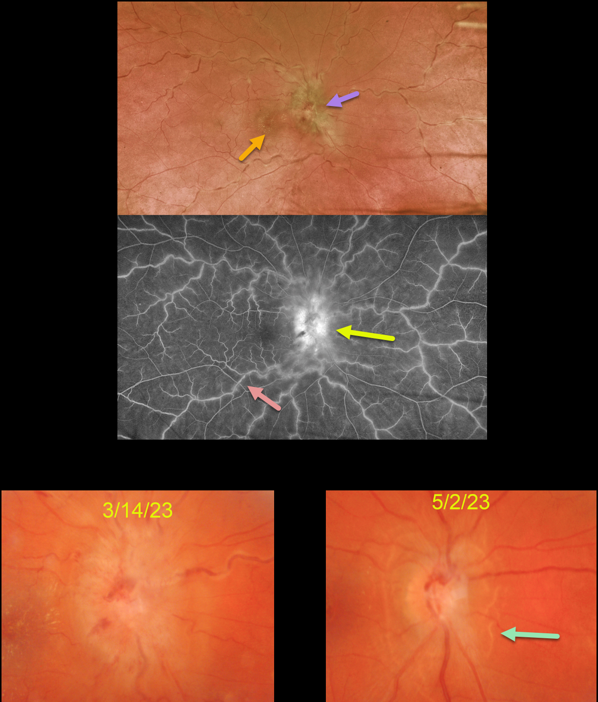
This 36YO female presented with unilateral vision loss in her left eye. She denied any systemic symptoms except for headaches for 3 months. Vision was 20/70 in her right eye and 20/25 in her left eye.
Optos color imaging shows severe optic nerve head edema, a foveal lipid star, dilated and tortuous vessels, and a variable frosted branch appearance to the retinal veins. Fluorescein angiography shows diffuse venous staining with optic nerve leakage.
Extensive blood work was positive for both Bartonella henselae (IgG 1:64) and Bartonella quintana (IgG 1:128), and negative for Bartonella IgM antibodies. Although she has cats at home, she denied cat scratches or exposure to cat fleas. She was referred to an infectious disease specialist who started her on doxycycline 100mg PO BID.
When she returned 6 weeks later, her headaches had resolved, and her vision was 20/80. The optic nerve swelling was dramatically improved with secondary peripapillary pigmentary changes.
Learning Points:
Bartonella henselae is the bacterium associated with cat scratch disease. It is transmitted from cat fleas carrying the bacterium or from flea feces that can be present on the cat’s claws/teeth, and transferred to humans from a cat scratch, lick, or bite.
Common systemic findings include fever, malaise, and progressive lymphadenopathy. These findings usually occur within 1-2 weeks of a cat scratch/bite.
Ocular effects primarily include neuroretinitis, with rare cases of Parinaud’s ocular glandular syndrome and, as in our patient’s case, chorioretinitis with vasculitis.



