

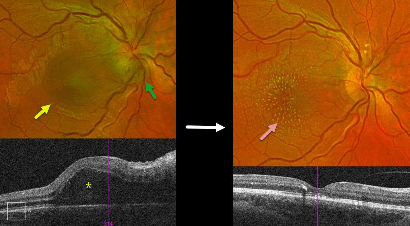
This 18YO female presented with a several-day history of vision loss in her right eye. Vision was 20/70 OD and 20/40 in her normal OS.
Optos color RG imaging shows inferior disc edema with a serous macular detachment. Triton swept-source OCT confirms the serous detachment extending towards the nerve.
Bartonella serology was positive, and she was placed on a 6-week course of oral antibiotics. Two months following presentation, vision improved to 20/40. The disc edema and serous detachment resolved, with the development of a lipid star.
Learning Points:
The diagnosis for a unilateral swollen nerve is extensive. However, it becomes much smaller when associated with acute inner retinal ischemia (not seen in our case) and, later, with lipid exudation within Henle’s layer (neuroretinitis), including cat-scratch (Bartonella) and syphilis. When no underlying cause is found, the entity is called Leber’s idiopathic stellate neuroretinitis



