

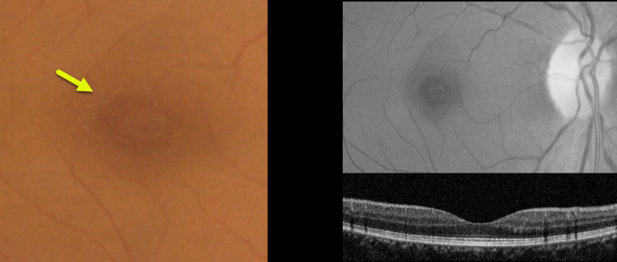
This 49YO female presented with asymptomatic macular changes. Vision was 20/30 OD and 20/40 OS. There was no past medical history and no family history of eye disease.
Color imaging shows tiny yellow perifoveal dots, which are somewhat better seen with red-free imaging. OCT scanning is unremarkable.
Learning Points:
Benign yellow dot maculopathy was reported as a new nonprogressive macular phenotype by Borman et al (Ophthalmology 2017;124:1004-1013).
Patients present with normal vision and asymptomatic tiny yellow foveal dots. These dots are hyperautofluorescent, but OCT scanning is generally normal. There is no known causative gene.



