

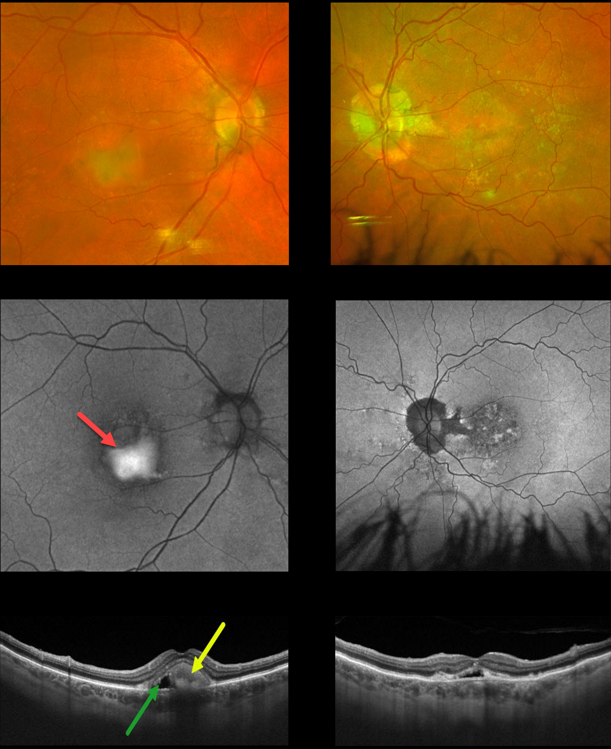
This 75YO male presented with chronic stable central blurriness. Vision was 20/150 OU.
Color photography of his right eye shows superior foveal pigment loss with inferior yellowish, vitelliform material, with more atrophic foveal changes in his left eye.
Fundus autofluorescence (FAF) shows the vitelliform material to be dramatically hyper-autofluorescent OD, with a more speckled hypo-FAF appearance OS.
Swept-source OCT scanning of his right eye shows hyperreflective inferior layering of the vitelliform material with a hyporeflective space more superiorly. Similar OCT findings are also seen in his left eye.
Our patient has progressed to a more atrophic, ‘scrambled egg’ appearance, most evident in his right eye. The remaining vitelliform material gravitates inferiorly, often leaving an optically empty, hyporeflective space between the RPE and outer retina. Eventually, the retina flattens over time with secondary atrophy, which is more pronounced in his left eye.
Learning Points:
Best disease is associated with a mutation in the BEST1 gene, which encodes the bestrophin-1 protein. Bestrophin-1, a calcium-activated chloride channel, is primarily found in the basolateral plasma membrane of the RPE.
BEST1 mutations cause a variety of phenotypes, including autosomal recessive bestrophinopathy, best vitelliform macular dystrophy, and autosomal dominant vitreoretinochoroidopathy.



