

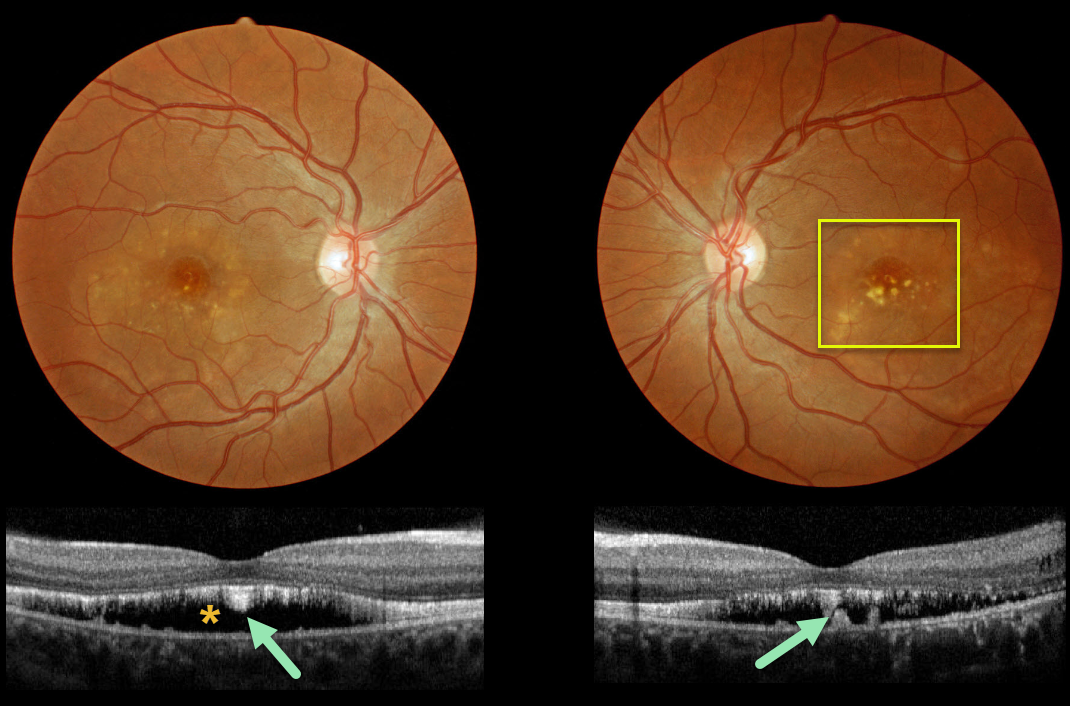
This patient has progressed to the more atrophic ‘scrambled egg’ appearance of Best’s disease.
OCT scanning shows the common hyporeflective subretinal space left behind after the yellow subretinal material absorbs, which often flattens over time.
Shaggy hyperreflective material, likely representing shed photoreceptors and residual vitelliform material, is noted along the posterior retinal surface and on top of the RPE.
Learning Points:
Best vitelliform macular dystrophy, or Best’s disease, is associated with a mutation in the BEST1 gene. This inherited disorder usually presents in childhood or early adulthood with a bilateral foveal “egg-yolk” appearance.



