

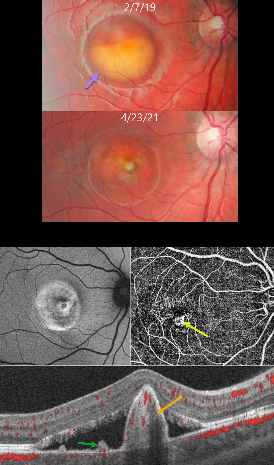
This healthy 11YO boy presented to us on 2/7/19 with asymptomatic retinal findings. Vision was 20/25 OD and 20/20 OS.
Color photography of his right eye shows a partially scrambled vitelliform lesion consistent with Best vitelliform macular dystrophy (BVMD). A smaller, scarred, and atrophic lesion was noted in his left eye (not shown).
He returned 2 years later without visual complaints. Vision was 20/40 OD. The vitelliform lesion in his right macula is atrophic, with a central depigmented nodule. Fundus autofluorescence (FAF) shows variable hyper- and hypo-FAF. OCTA en face shows a central flow signal. OCTA B-scan shows a nodular subretinal hyperreflective pillar containing flow signals, subretinal fluid, and vitelliform material layered on the RPE and lining the outer retina. The vision and OCT findings did not change following 2 intravitreal Lucentis injections, so observation was recommended. We continue to follow him with stable findings 5 years later.
Learning Points:
BVMD is associated with a mutation in the BEST1 gene, which encodes the bestrophin-1 protein. Bestrophin-1, a calcium-activated chloride channel, is primarily found in the basolateral plasma membrane of the RPE. BEST1 mutations cause a variety of phenotypes, including autosomal recessive bestrophinopathy, best vitelliform macular dystrophy, and autosomal dominant vitreoretinochoroidopathy.
Secondary macular neovascularization (MNV) develops in up to half of eyes, depending on the imaging modality used, and can develop at any stage of the disease. MNV exists in two forms: the less common exudative form, which presents with acute vision loss, subretinal hemorrhage, and intraretinal/subretinal fluid, and the more common non-exudative form seen in more advanced (vitelliruptive and atrophic) stages (Parodi et al, Invest Ophthalmol Vis Sci 2020;61(6);61).
These subclinical MNVs are best detected with swept-source OCTA. Structural OCT findings that suggest non-exudative MNV include focal choroidal excavations and nodular subretinal pillars (as in our patient). Exudative MNV responds well to standard anti-VEGF injections, whereas non-exudative lesions tend to remain stable without treatment.



