

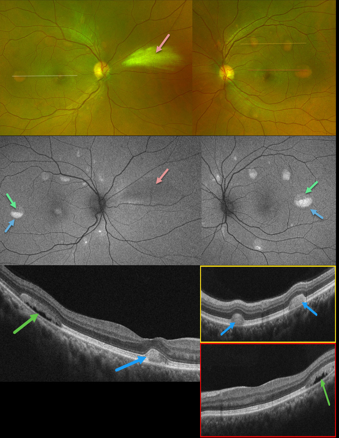
This 39YO male presented with asymptomatic retinal lesions. Family history was negative. Vision was 20/20 OU.
Optos color RG imaging shows bilateral partially scrambled vitelliform lesions in each posterior pole. On OCT, the superior, scrambled portion of these lesions is hyporeflective, and the inferior, yellow portion is hyperreflective. On fundus autofluorescence (FAF), the superior portions are hypo-FAF, and the inferior vitelliform material is hyper-FAF. The incidental area of the nasal myelinated nerve fiber layer OD is hypo-FAF from blocking the underlying RPE.
Learning Points:
Vitelliform lesions are characterized by the accumulation of yellow subretinal material thought to be caused by impaired metabolism of photoreceptors or retinal pigment epithelium. They can be associated with a wide variety of retinal disorders, including Best vitelliform macular dystrophy (BVD, our patient), adult-onset foveomacular dystrophy, acute exudative polymorphous vitelliform maculopathy, as well as paraneoplastic, toxic, tractional, and degenerative etiologies (Iovino et al, Surv Ophthalmology 2023;68:361-379).
BVD is usually transmitted as an autosomal-dominant disorder due to a mutation in the BEST1 gene, which encodes bestrophin-1. Bestrophin-1, a calcium-activated chloride channel, is primarily found in the basolateral plasma membrane of the RPE. Unfortunately, our patient denied genetic testing due to cost.



