

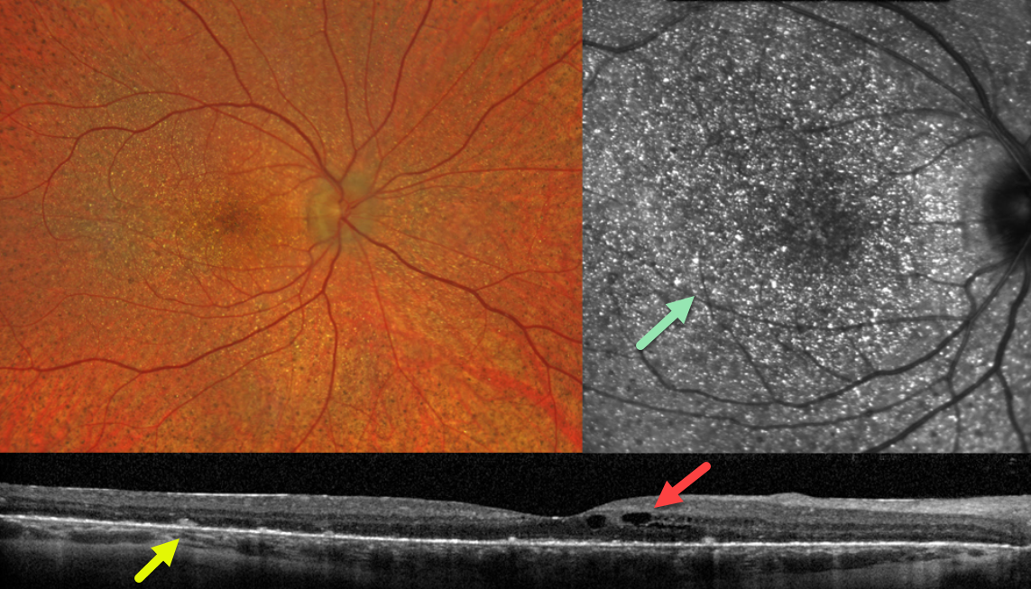
This 28YO female has a known history of Bietti crystalline dystrophy and was without new visual complaints. Vision was 20/50 OD and 20/40 OS.
Fundus imaging of her right eye shows extensive subretinal crystalline deposits primarily in the posterior poles, with tiny subretinal pigment clumps more peripherally.
OCT scanning shows hyperreflective dots overlying the RPE-Bruch’s membrane complex, with variable outer retinal and RPE loss. Cystic changes are noted in the inner nuclear layer.
En face OCT through the outer retina reveals innumerable hyperreflective crystalline deposits. Identical findings were noted in her left eye.
Learning Points:
Bietti crystalline dystrophy is an autosomal recessive ocular disorder that affects both the anterior and posterior segments. Clinical features include crystalline deposits in the retina and cornea, and, rarely, in the crystalline lens, as well as retinal pigment epithelial clumping and atrophy.
Mutations in CYP4V2, which cause dysfunctional lipid metabolism, have been implicated in its pathogenesis. While there are no definite treatments, anti-VEGF therapy can be given for secondary macular neovascularization.
See Saatci et al for a recent review (Clinical Ophthalmology 2023;17:953-967).



