

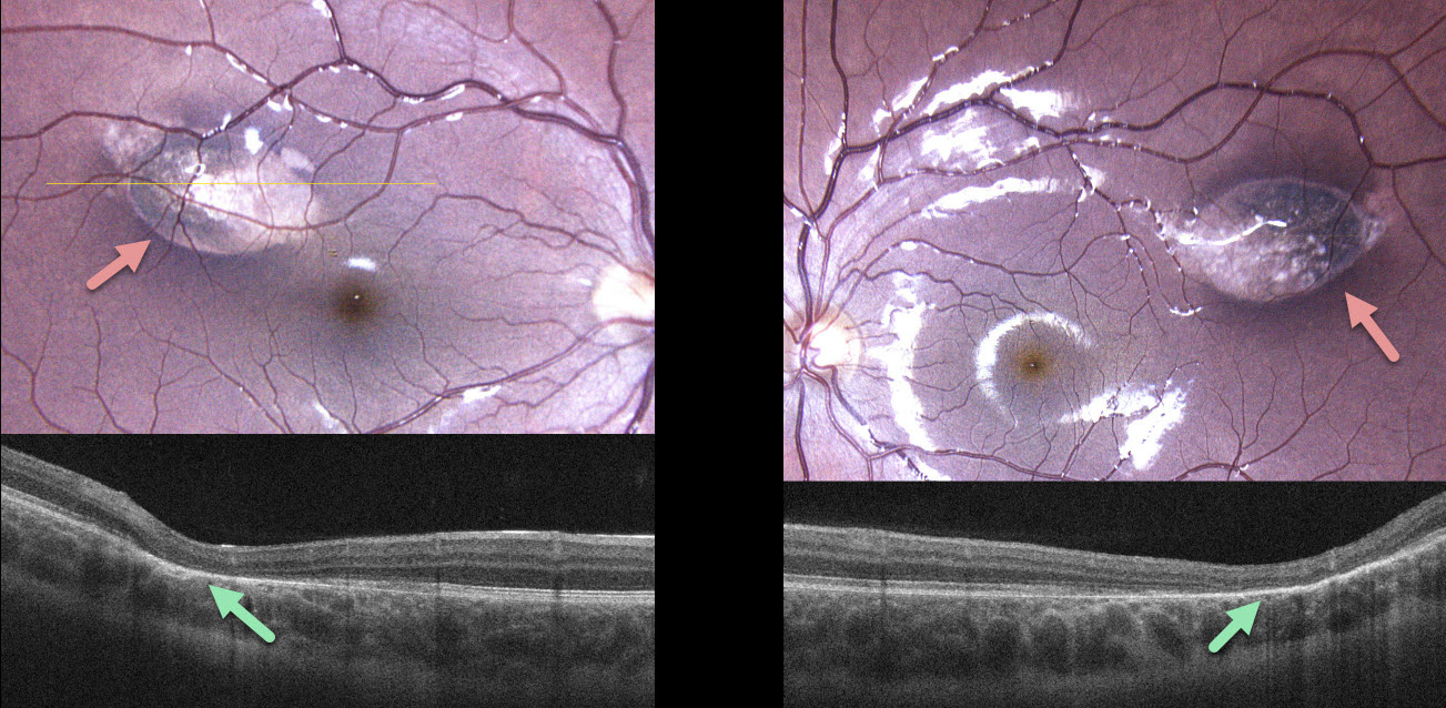
This 10YO boy was referred for asymptomatic retinal lesions. He was adopted, so the family history was unknown. The patient and guardian reported a longstanding history of persistent diarrhea that at times required hospitalization. Vision was 20/20 bilaterally.
Color photography shows bilateral torpedo-shaped areas of depigmentation in each superotemporal macula. The lesions were hypo-autofluorescent (not shown). OCT scanning shows outer retinal atrophy through each lesion.
Learning Points:
Torpedo maculopathy was originally described by Gass as a solitary hypopigmented nevus of the RPE (Arch Ophthalmology 1992;110:1358-1359). These benign lesions are teardrop-shaped and are almost always located in the horizontal meridian in the temporal macula. Vision is almost always normal. There is a small risk of macular neovascularization.
The lesions are virtually always unilateral, and we found only a single case report of bilateral lesions in a PubMed search (Richez et al., J Fr Ophthalmol 2010;33:296). Given our patient’s GI symptoms and bilaterality, genetic testing was recommended, but he was unfortunately immediately lost to follow-up.



