

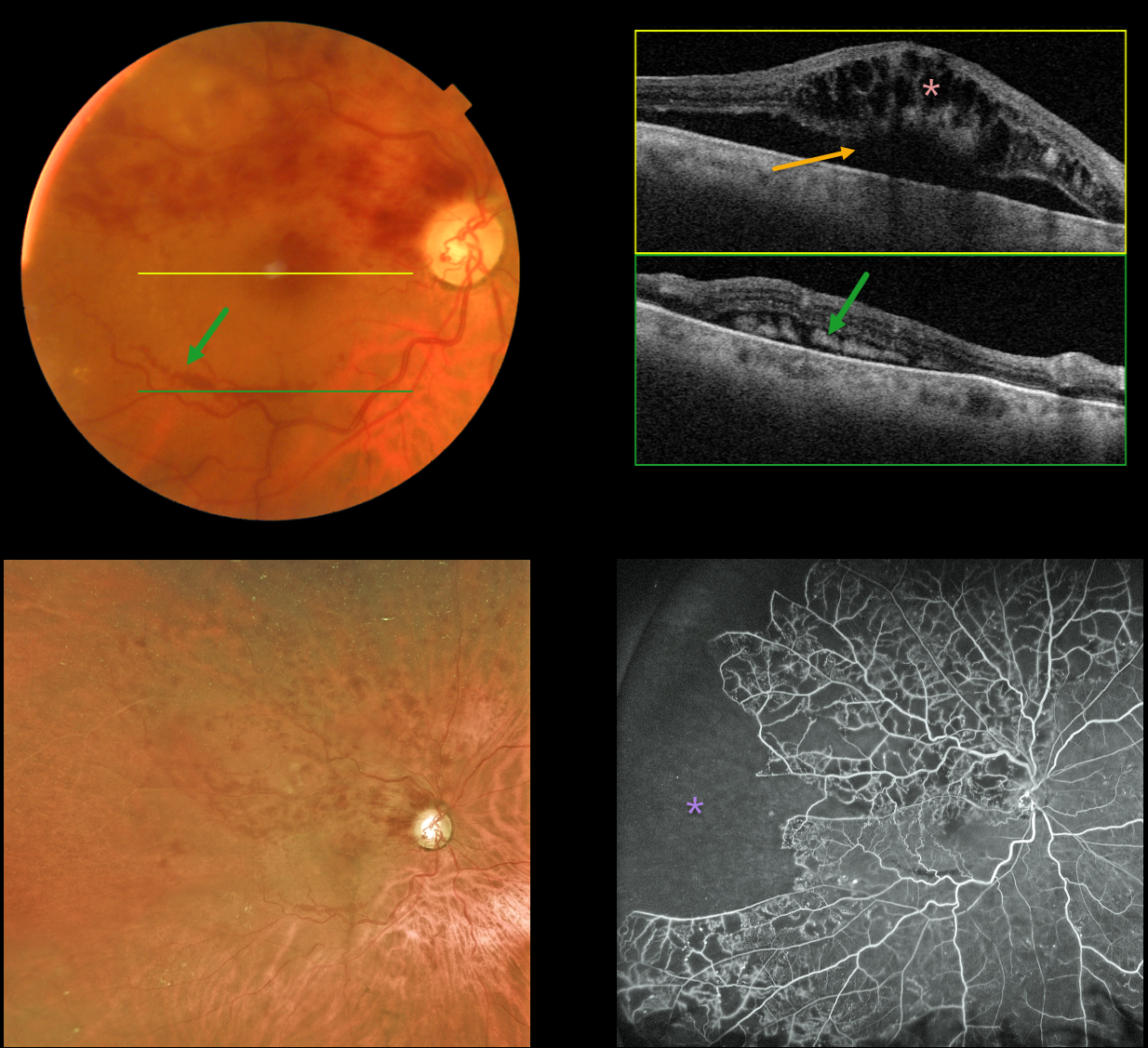
This 79YO male presented with recent vision loss of 20/400 in his right eye from a fresh branch retinal vein occlusion (BRVO). Layered subretinal blood is noted in the inferior macula.
Swept-source OCT through the macular center shows marked cystoid macular edema with underlying subretinal fluid. OCT through the inferior macular subretinal blood shows subretinal hyperreflectivity. Optos fluorescein angiography shows marked peripheral ischemia without neovascularization.
Learning Points:
Layered preretinal blood is a common occurrence with proliferative retinopathies, including proliferative diabetic retinopathy and BRVO. The retinal neovascularization grows from the retinal surface onto the back of the posterior hyaloid, and blood accumulates in the subhyaloid space.
The appearance of layered inferior macula blood in our patient is a ‘fake-out’ because the blood was neither neovascular nor preretinal. Blood, likely due to hydrostatic pressure, was forced into the subretinal space, and the subretinal fluid allowed it to settle inferiorly.
The blood and fluid completely resolved following several monthly intravitreal anti-VEGF injections.



