

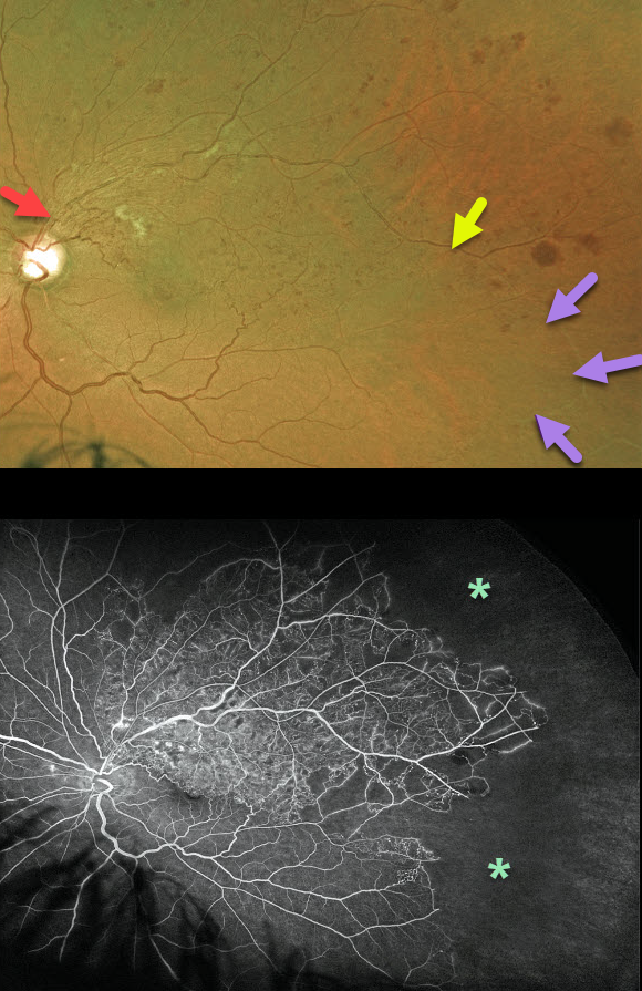
This is a 54YO male presented with a major branch retinal vein occlusion (BRVO). Vision was 20/80, and Eylea injections were started for associated macular edema.
The Optos color image shows a major superotemporal BRVO with moderate retinal hemorrhages, some nerve fiber layer infarcts, and collateral vessels. The involved retinal vein is red in the midperiphery. However, it becomes white as it continues into the inferotemporal periphery.
Optos fluorescein angiography shows extensive peripheral capillary dropout.
Usually, one would expect a BRVO to involve one quadrant (retinal vessels virtually always respect the horizontal meridian).
The occluded vein in our patient most likely represents an atypical retinal venous malformation (RVM), which is a congenital anomalous vessel that, by definition, crosses the horizontal.
Learning Points:
Retinal venous malformations were originally described by Gary Brown et al as congenital retinal macrovessels (Arch Ophthalmology 1982;100:1430-1436). These vessels are usually located in the macular region and are virtually always venous.
These lesions are now called retinal venous malformations (RVMs) since about one-quarter of patients have associated venous anomalies in the brain (Pichi et al., JAMA Ophthalmol 2018;136;372-379).



