

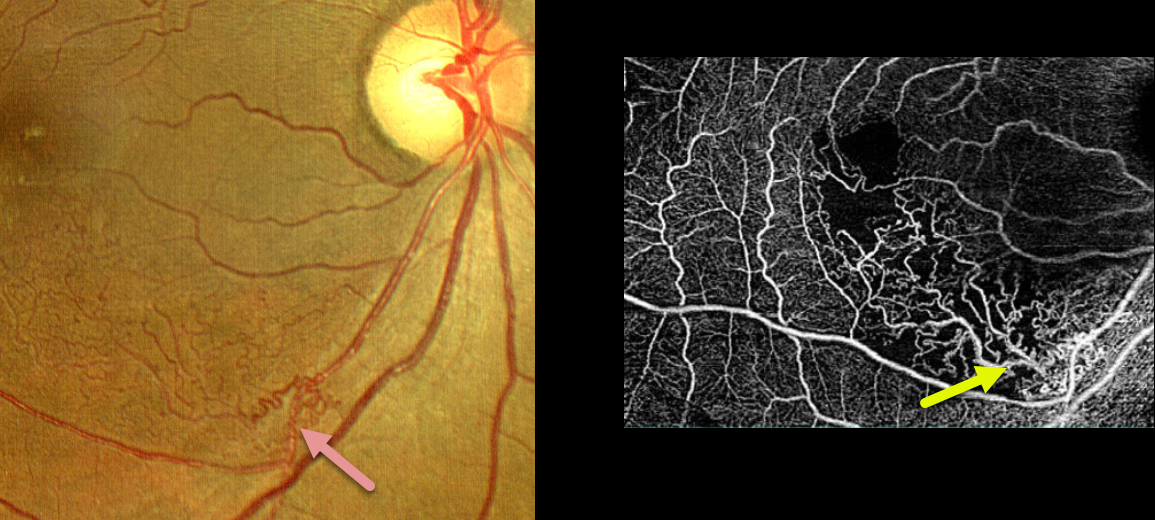
This 53YO female presented with 20/80 vision in her right eye from a macular branch retinal vein occlusion (BRVO) with macular edema. The edema resolved following a single injection of a Lucentis biosimilar. She was then lost to follow-up for 8 months, when she returned with 20/20 vision.
Extensive inferonasal macular collateral vessels are seen on fundus photo and OCTA. OCT showed no edema (not shown).
Learning Points:
Collateral vessels are dilated, pre-existing vessels that bypass an occlusion. These are most commonly seen at the posterior pole following retinal venous occlusions, but can also occur at the optic disc with optic nerve meningiomas. The endothelial tight junctions are intact, so they don’t leak on fluorescein angiography. This helps differentiate them from neovascularization, which does leak.
Collaterals most commonly form in the deep vascular plexus, which represents the primary venous drainage for the inner retina (Freund et al, JAMA Ophthalmology 2018;1262-1270).



