

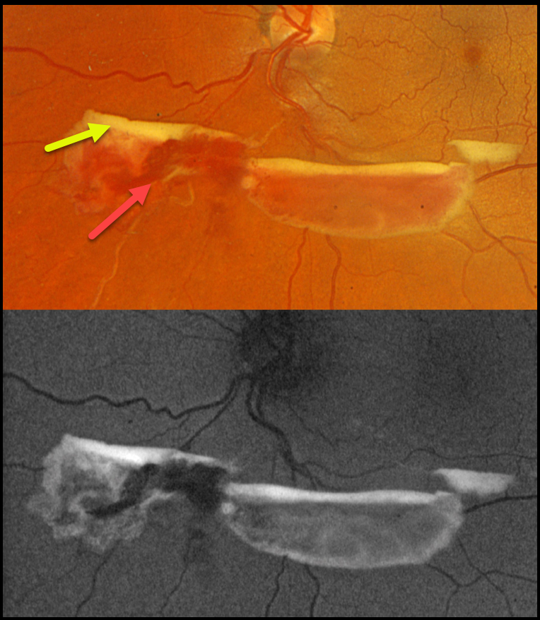
This 48YO female was referred for asymptomatic retinal findings. Vision was 20/20 in the normal OD and 20/20 OS.
Optos color RGB imaging shows red and yellow preretinal blood just inferior to the nerve due to an ischemic extramacular inferonasal branch retinal vein occlusion (BRVO). On fundus autofluorescence (FAF), the red blood is hypo-FAF, and the devitalized yellow blood is markedly hyper-FAF. Intravitreal Avastin was given, and scatter laser was scheduled, although she was immediately lost to follow-up.
Learning Points:
Porphyrins are organic molecules that are part of the heme molecule in hemoglobin. When these compounds break down, the devitalized blood appears yellow and becomes intensely hyperautofluorescent (Bloom and Spaide, Retinal Cases & Brief Reports 2022;16:401-402).



