

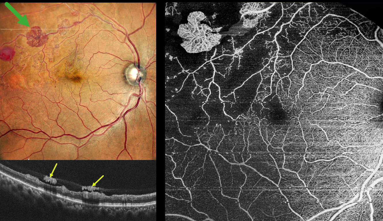
This 55YO hypertensive male presented for a routine eye examination. He gave a history of a resolved superotemporal branch retinal vein occlusion (BRVO) with macular edema in his right eye that was successfully treated with a single anti-VEGF injection in 2022. Vision was 20/15 OD and 20/15 in his normal OS.
Pseudocolor SLO imaging shows an ischemic superotemporal BRVO with several fronds of active red and variably fibrosed retinal neovascularization. A round deep retinal hemorrhage is noted distally. On OCT, the neovascularization appears as hyperreflective plaques along the posterior edge of the partially detached posterior hyaloid. OCT angiography confirms diffuse capillary nonperfusion, a relatively preserved foveal avascular zone, and flow signals within the areas of neovascularization.
Learning Points:
An attached vitreous provides the scaffold for neovascularization to grow onto and spread. Retinal neovascularization grows from the inner retinal surface and proliferates along the outer posterior hyaloid face (Vaz-Pereira et al, Retina 2017;37:1287-1296). This tethering to a partially detached and contracting hyaloid is what causes tractional retinal detachment and vitreous hemorrhage in proliferative retinopathies.
In 1986, the Branch Vein Occlusion Study Group reported that scatter laser decreased the risk of vitreous hemorrhage from retinal neovascularization in BRVO and suggested that laser should be applied once neovascularization develops (Arch Ophthalmol 1986;104:34-41). Almost 40 years later, scatter laser remains the gold standard, although some may prefer to stabilize neovascularization with anti-VEGF therapy before laser. An anti-VEGF injection, followed by a superotemporal scatter laser, was therefore recommended for our patient.



