

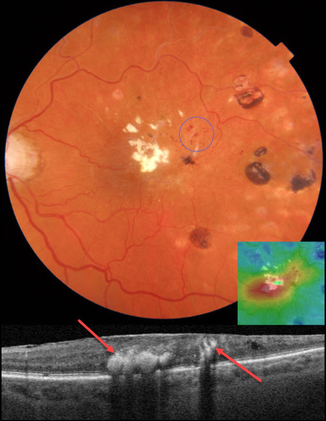
This 83YO female presented with recent vision loss in her left eye. There was a prior history of a branch retinal vein occlusion (BRVO), for which she received scatter laser elsewhere 8 years earlier. Vision was 20/60 OS.
Color photography shows superotemporal macular telangiectasia with likely retinal venous macroaneurysms (RVMs). Fairly dense lipid is noted in the fovea along with temporal laser scarring.
On OCT scanning, the lipid is markedly hyperreflective with posterior shadowing. There is central macular edema on the thickness map. Focal thermal laser was recommended to the RVMs.
Learning Points:
In 1990, Cousins et al reported that RVMs can be an additional finding in occlusive venous disease, always in association with collateral vessels or capillary or arterial macroaneurysms (AJO 1990;109:567-570).
These vascular abnormalities tend to have a higher rate of intraretinal lipid (40%) but are less likely to affect central vision since they are usually outside the macula. They tend to have a higher rate of developing retinal neovascularization (32% of eyes).



