

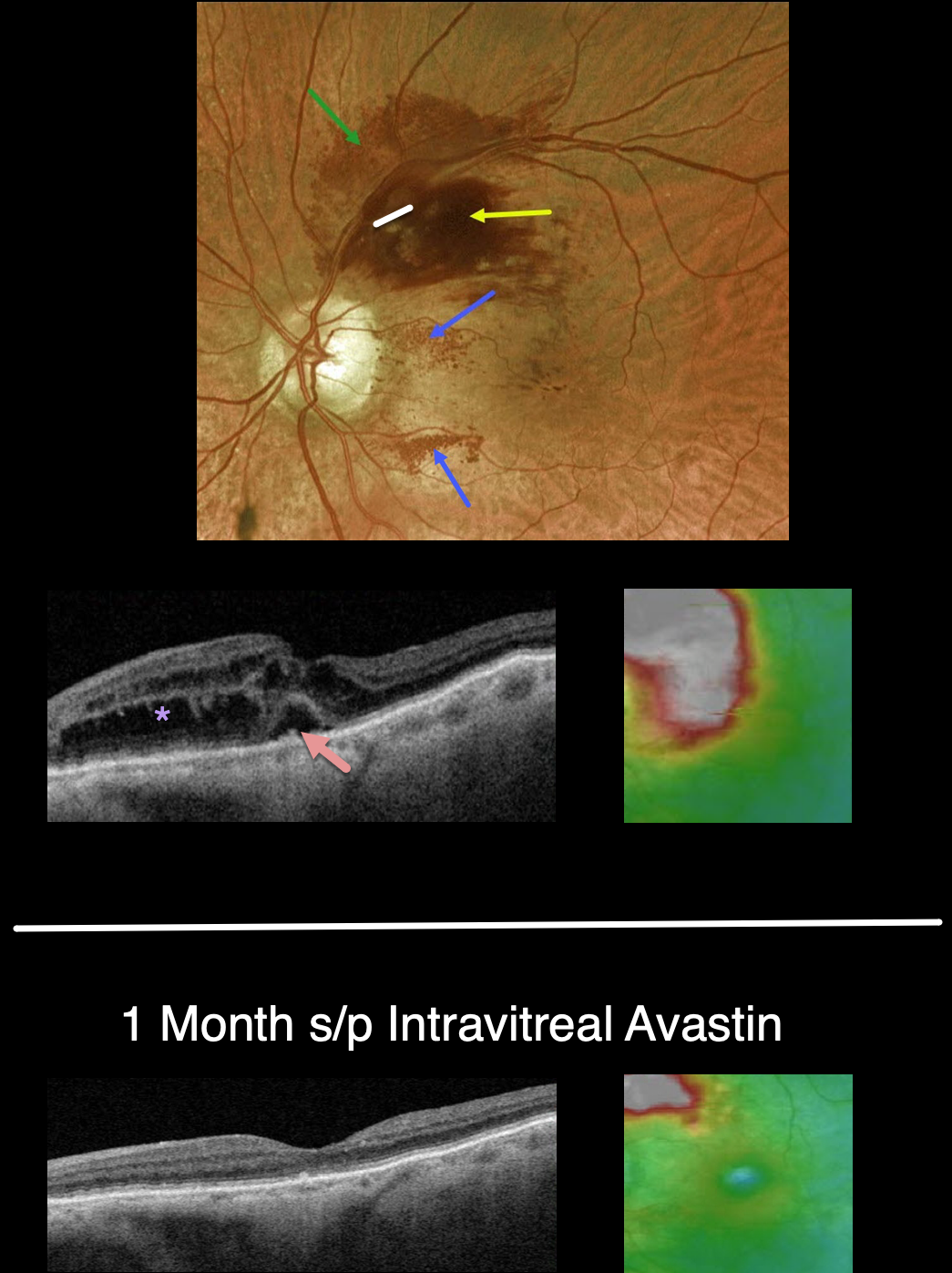
This 88YO female presented with recent vision loss in her right eye. Vision was 20/50.
Optos color imaging shows an unusual appearing superotemporal branch retinal vein occlusion (BRVO). Confluent inner retinal hemorrhages are noted in the superonasal macula, with milder deep retinal blood more superiorly. Unusual dots of intraretinal blood are also noted nasally and inferonasally.
Swept-source OCT shows moderately severe central and superonasal macular edema with some foveal subretinal fluid.
An intravitreal Avastin injection was given. One month later, vision improved to 20/30. The retinal hemorrhages dramatically improved. OCT scanning shows complete resolution of all central fluid, with some residual distal superonasal edema.



