

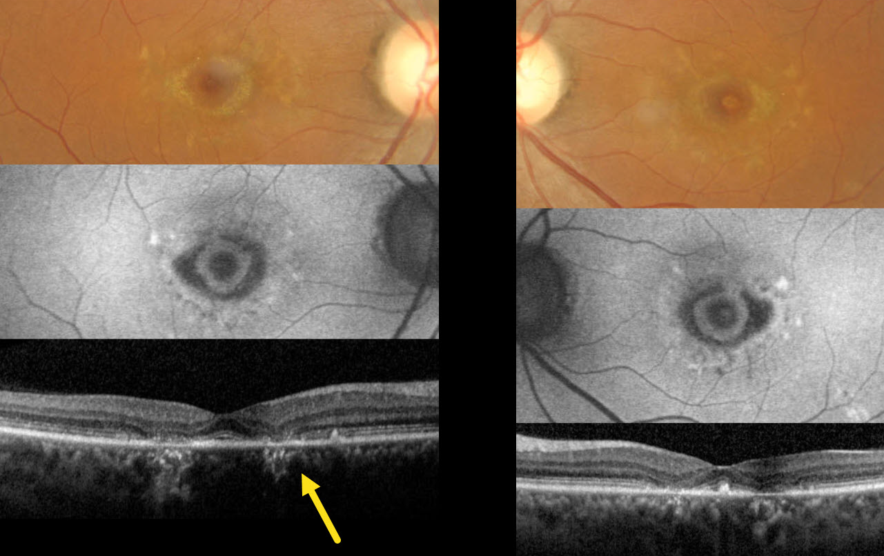
This 40YO female presented with 5 years of stable, mild, distorted vision bilaterally. There was no family history of eye disease. Vision was stable at 20/20 OU.
Color imaging shows bilaterally symmetric bullseye lesions with surrounding subretinal yellow macular flecks. On fundus autofluorescence (FAF), these lesions are hypo-FAF with an inner hyper-FAF ring. Pericentral outer retinal atrophy is noted on OCT.
The funduscopic appearance is most consistent with ABCA4 disease, including Stargardt disease and fundus flavimaculatus. The patient declined genetic testing due to cost concerns, and continued observation was recommended.



