

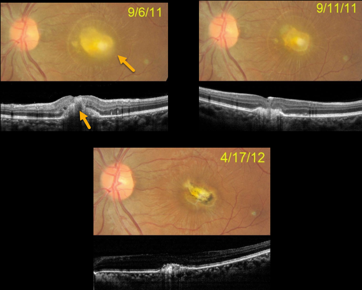
In 2011, this 27YO female was recently hospitalized and treated with intravenous amphotericin. Upon discharge, she was examined for vision loss in her left eye. Vision was hand motion.
Color photography shows a thickened yellow subfoveal lesion that extends through the full-thickness retina on OCT. One week later, vision improved to counting fingers, and the lesion is flattening and contracting. Seven months later, a small contracted foveal scar remains, with 4/60 vision. She was subsequently lost to follow-up.
Learning Points:
Ocular candidiasis is characterized by single or multiple focal yellow-white chorioretinal lesions with overlying vitritis, vitreous haze, vitreous abscess (fluff balls or string of pearls), and other more generalized presentations of infection and inflammation, such as endophthalmitis, hypopyon, scleritis, exudates, and anterior chamber cells. Ocular candidiasis can be caused by exogenous or endogenous sources. Exogenous routes include trauma and inoculation from surgical procedures. Endogenous ocular candidiasis arises from hematogenous seeding of chorioretinal blood vessels via fungemia, indwelling catheters, a severely compromised immune system, or broad-spectrum antibiotic abuse, etc.
Treatment includes removal of the inciting source of candidiasis (i.e., indwelling catheter) and systemic therapy with possible intravitreal antifungal therapy.
Routine ophthalmic screening for known systemic candida sepsis is not recommended unless there are suggestive signs or symptoms for ocular involvement (see Breazzano et al, Ophthalmology 2011;129:73-76).



