

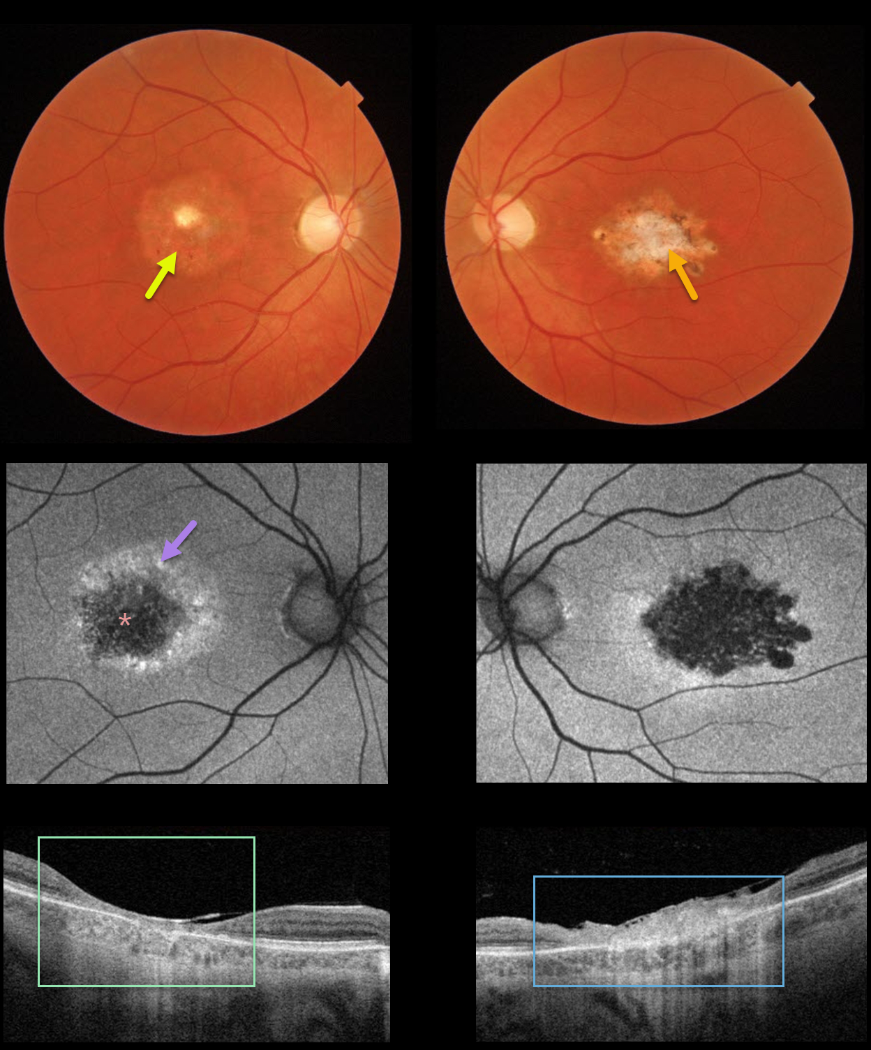
This 40YO female gave a 4-5 year history of gradual bilateral vision loss. Vision was 20/70 OD and 20/200 OS. There was no family history of retinal disease.
Color imaging shows a round area of central pigment loss OD and a more irregular area of atrophic pigmentary changes OS.
Optos fundus autofluorescence (FAF) showed central hypo-FAF with surrounding hyper-FAF.
Swept-source OCT showed a markedly thinned and disorganized central macula OD, with a more thickened, hyperreflective, disorganized central macula OS.
Genetic testing revealed a heterozygous pathogenic mutation in IQCB1, a defect commonly associated with autosomal recessive Senior-Løken Syndrome. We felt the clinical picture was most consistent with central areolar choroidal dystrophy (CACD).
Learning Points:
CACD is usually autosomal dominant and genetically heterogeneous, caused by mutations in the peripherin/RDS (PRPH2) gene. The current genetic testing panel results, as this case illustrates, must still be approached with caution (see Rodriguez-Munoz et al, Retina 2021;41:1966-1975).
See Smailhodzic et al Invest Ophthalmol Vis Sci 2011;52;8908-8918 for how to differentiate CACD from age-related macular degeneration best clinically.



