

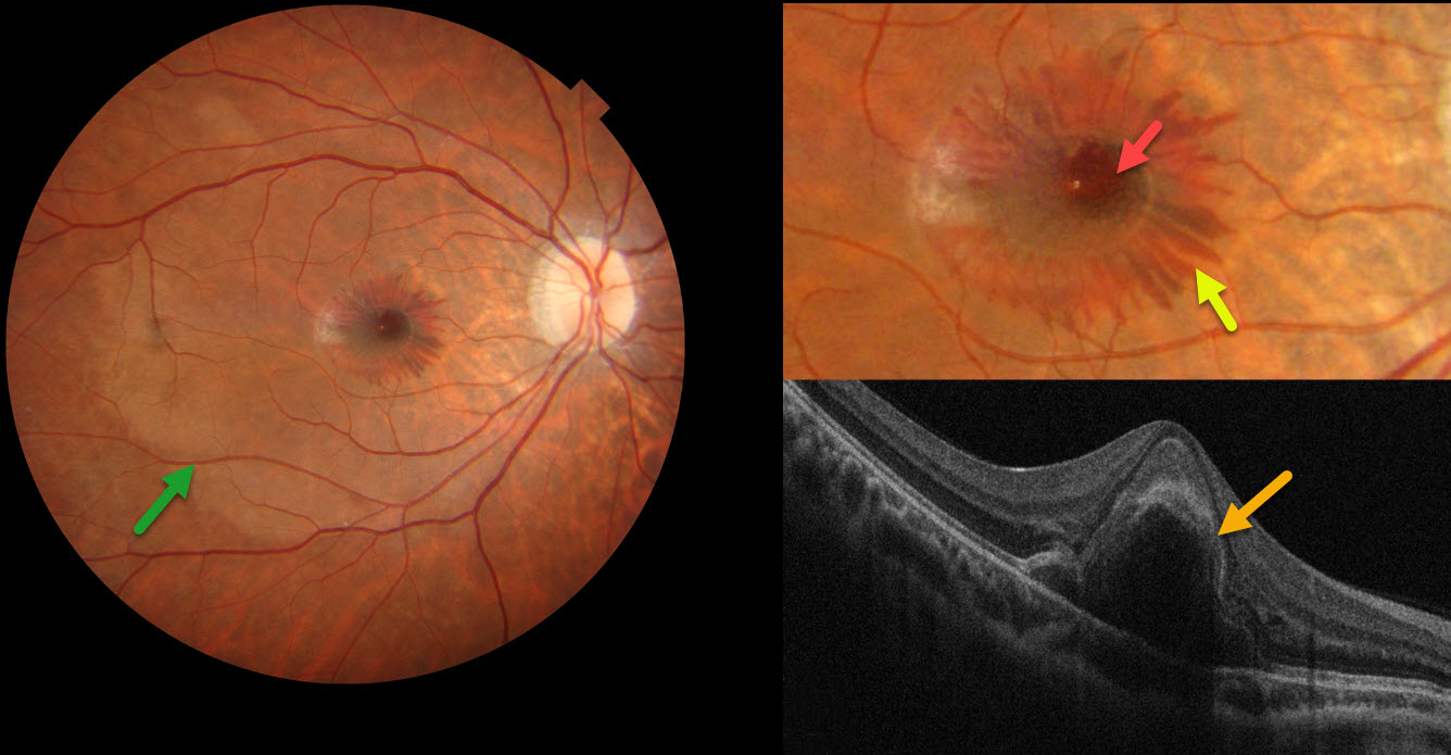
This 24YO male woke with distorted vision in his right eye the day he presented to our clinic. Over the next 2 hours, a central spot appeared in his vision. He recalled an active tennis game the previous day but denied any direct ocular trauma. Vision was 20/200 OD and 20/25 in his normal OS.
Color photography shows central foveal blood, surrounding radiating outer plexiform (OPL) blood, and a deep vs subretinal whitish discoloration of the macula. On OCT, the central blood is located in the outer retina.
Learning Points:
Radiating deep macular hemorrhages are seen in multiple conditions, including Valsalva and trauma. Their unique radiating appearance is caused by blood accumulating in Henle’s layer. Kon Graversen et al described hemorrhagic unilateral retinopathy, an idiopathic disorder with a similar appearance (Retina 2014;34:483-489).
Ramthohul et al recently described a central bouquet hemorrhage with a round intraretinal blood spot surrounded by these OPL hemorrhages (Retina 2024;44:551-557). Various etiologies included neovascular AMD, myopic lacquer cracks, macular telangiectasia, and trauma associated with angioid streaks. Despite the macular changes consistent with commotio retinae, our patient adamantly denied ocular trauma.



