

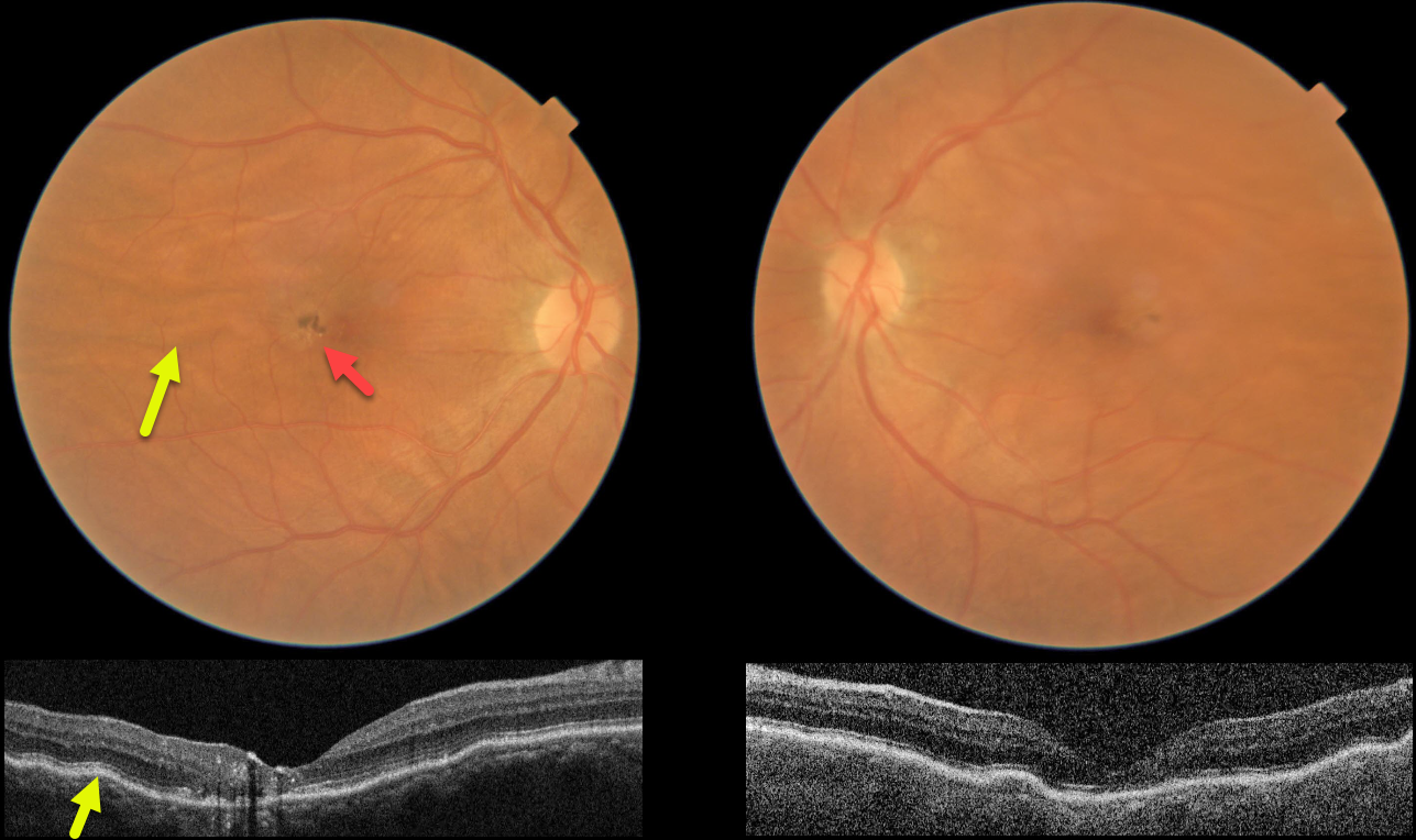
This 61YO female presented without a visual complaint. She was a +4 diopter hyperope bilaterally. Vision was 20/200 OD and 20/20 OS.
Color imaging and OCT show bilateral chorioretinal folds, which are particularly prominent in her right eye. Foveal pigmentary changes are noted in the central right macula, with OCT disorganization of the central macular layers and hyperreflective foci. Observation was recommended.
Learning Points:
Chorioretinal folds appear clinically as often subtle horizontal and oblique subretinal folds. There are extensive intra- and extra-ocular causes for chorioretinal folds. The typical findings in idiopathic chorioretinal folds are a mild/moderate hyperopic prescription and bilateral, symmetric folds. The folds are thought to result from flattening of the macular sclera.
When unilateral, an MRI is often indicated to rule out an orbital mass. Typical idiopathic chorioretinal folds require no workup and usually cause no symptoms.
In our practice, we have seen several patients with idiopathic chorioretinal folds and waxing/waning subretinal fluid (SRF), possibly as part of the pachychoroid spectrum.
Cohen et al. also reported a small series of patients with hyperopic chorioretinal folds, central serous retinopathy, and pachychoroid (Retinal Cases 2022;16:242-245). We therefore feel the pigmentary changes noted in our patient’s right eye most likely resulted from spontaneously resolved SRF.



