

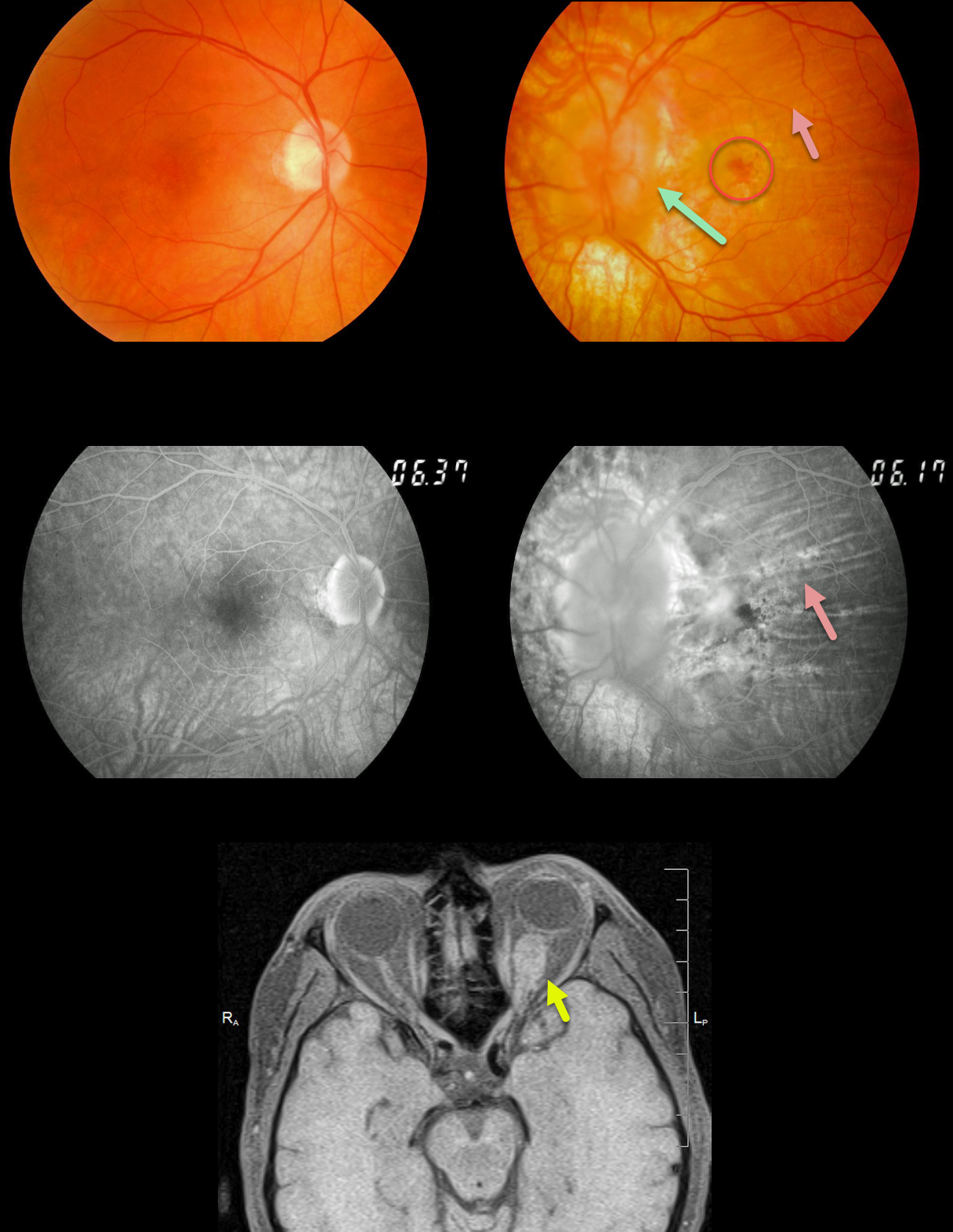
This patient presented with a history of a left optic nerve meningioma that had been followed for years. The left nerve is markedly swollen.
Extensive chorioretinal folds are present and best seen on angiography. A chronic subfoveal macular neovascularization (MNV) is noted, which we are observing since the lesion and vision remain stable at 20/400.
MRI shows the optic nerve meningioma; this is also being observed since surgical removal of this benign tumor would cause total blindness.
Learning Points:
Chorioretinal folds are usually idiopathic and bilateral, most commonly from mild hyperopia and flattening of the macular sclera.
When folds present unilaterally, as in our patient, an MRI of the orbit is recommended to rule out orbit pathology.



