

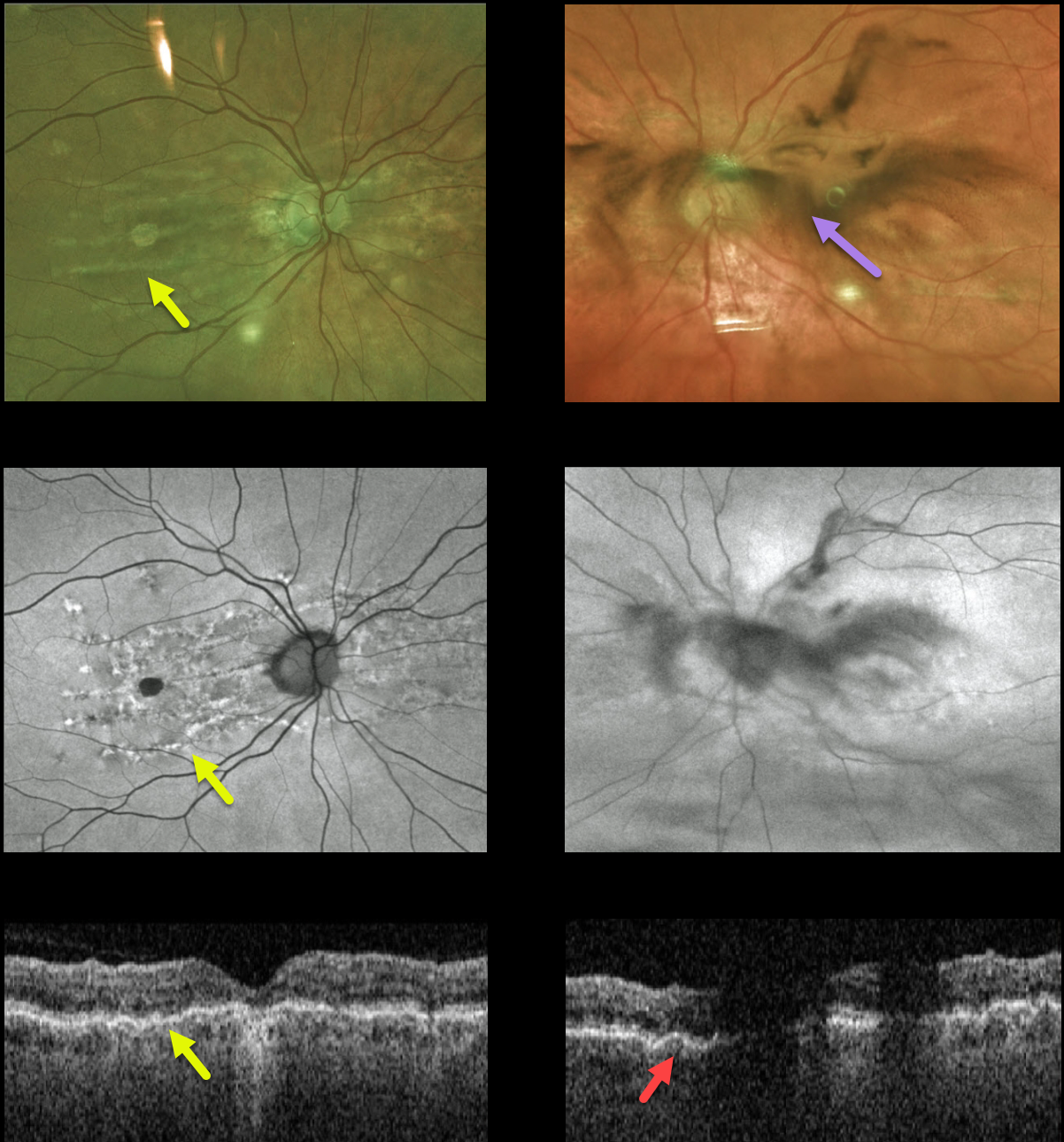
This 80YO female presented with a 2-day history of floaters and vision loss in her left eye. She had mild hyperopia prior to cataract surgery 5 years earlier. She had a history of diet-controlled type 2 diabetes. Vision was 20/100 OD and 20/30 OS.
Fundus Optos color imaging, fundus autofluorescence, and OCT of her right eye show changes associated with chronic chorioretinal folds. Similar imaging of her left eye is hazy due to a vitreous hemorrhage, but chorioretinal folds are also noted on the OCT B-scan.
There were no signs of diabetic retinopathy clinically or on fluorescein angiography (not shown), and no signs of retinal breaks or detachment peripherally. She was asked to sleep with her head elevated, and we are following her closely.
Learning Points:
Chorioretinal folds appear clinically as often subtle horizontal and oblique subretinal folds. There are extensive intra- and extra-ocular causes for chorioretinal folds. When unilateral, an MRI is often indicated to rule out an orbital mass. Typical idiopathic chorioretinal folds require no workup and usually cause no symptoms.
Our patient shows typical findings of idiopathic chorioretinal folds, including mild hyperopia and bilateral, symmetric folds. The folds are thought to result from flattening of the macular sclera.



