

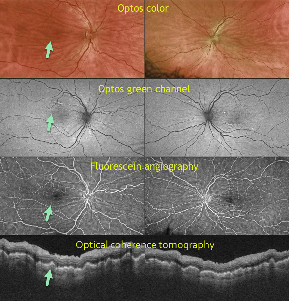
This 50-year-old male is a 6-diopter hyperope. Optos color, Optos green channel, fluorescein angiography (FA), and OCT reveal prominent chorioretinal folds.
Learning Points:
Chorioretinal folds appear clinically as often subtle horizontal and oblique subretinal folds. There are extensive intra- and extra-ocular causes for chorioretinal folds.
Our patient shows typical findings of idiopathic chorioretinal folds, including mild hyperopia and bilateral, symmetric folds. The folds are thought to result from flattening of the macular sclera.
When unilateral, an MRI is often indicated to rule out an orbital mass. Typical idiopathic chorioretinal folds require no workup and usually cause no symptoms.



