

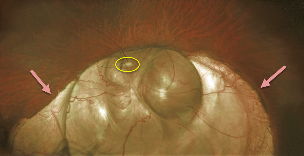
This patient has a large choroidal coloboma extending through the left optic nerve and macula. It’s so extensive that it hides the optic nerve.
These arise from incomplete closure of the embryonic fissure during the second month of embryonic development. Patients are at lifelong risk for rhegmatogenous retinal detachment due to breaks that occur in the overlying thinned retina.
Learning Points:
Defects in closure have a wide range of results such as microphthalmia, anophthalmia, iris coloboma, ciliary body coloboma, choroid coloboma, etc.
Colobomas of the iris or ciliary body result from failure of anterior closure while choroid, retina and optic nerve result from failure of posterior closure.
A staphyloma by definition is an outpouching of the entire eye wall. Choroidal coloboma, always involves a choroidal defect, but may or may not also involve the sclera (as in this case).



