

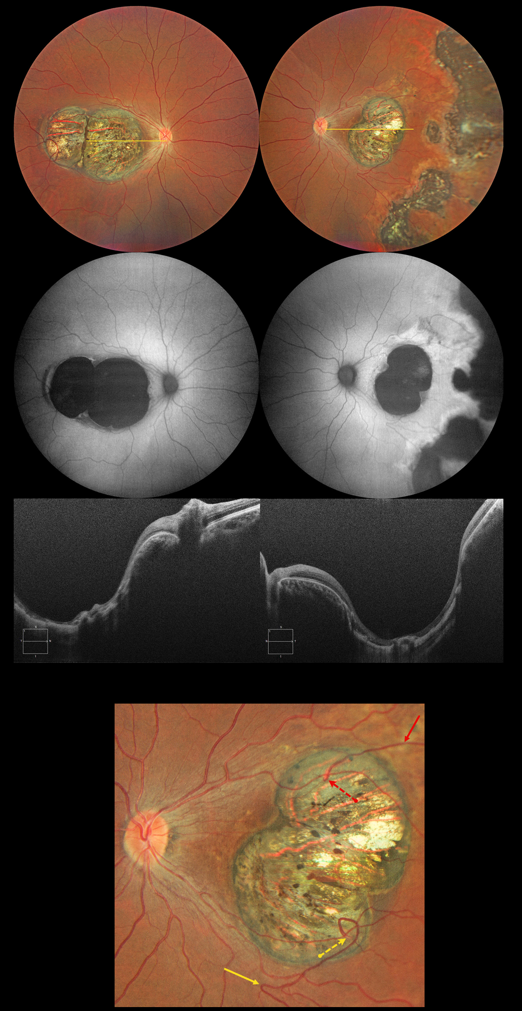
This 32YO female has a history of congenital nystagmus and poor vision since birth. Vision was 20/130 OU.
Color photography shows bilateral macular colobomas with more peripheral multifocal variably pigmented colobomatous scarring in her left eye. These lesions are hypoautofluorescent. OCT scanning through the macular lesions shows thinned hyperreflective retina draped across the posteriorly bowed sclera.
The right macular coloboma also contains several retinal choroidal anastomoses. The major inferotemporal vein acutely bends superotemporally to enter the coloboma and drain into the choroidal circulation. A superotemporal vein also enters the coloboma to drain into a visible choroidal vortex.
Learning Points:
Choroidal colobomas are most commonly caused by failure of the optic vesicle and choroidal fissure to close during embryonic development and are usually found inferonasally. They can present as solitary or multiple lesions. The differential diagnosis for congenital macular coloboma also includes post inflammatory lesions (toxoplasmosis, cytomegalovirus, and zika) and retinal dystrophies including North Carolina macular dystrophy. The retina within the coloboma is variably thinned, which increases the risk for rhegmatogenous retinal detachment.
Under normal conditions the retinal and choroidal circulations remain separate and distinct. Rarely, the two circulations directly connect with each other through a chorioretinal anastomosis. These can occur in chorioretinal scars (typically from inactive toxoplasmosis lesions), in macular telangiectasia, and in disciform scars.



