

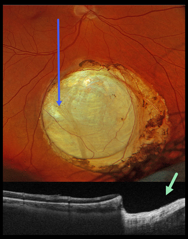
This healthy 48YO male was referred for an asymptomatic lesion in his right eye. Vision was 20/20 OD and 20/20 in his healthy OS.
Optos color RGB imaging shows an oval choroidal coloboma in the inferior midperiphery. Variably pigmented chorioretinal atrophy surrounds the lesion. Triton swept-source OCT shows a fairly abrupt and near-complete loss of the neurosensory retina within the coloboma.
Learning Points:
Choroidal colobomas are most often due to failure of the optic vesicle and choroidal fissure to close during fetal development. Lesions like this may also be from other causes, including toxoplasmosis. Choroidal coloboma can present as solitary or multiple lesions. Visual prognosis depends on the location and involvement of the optic nerve and macula.



