

This 12YO female complained of several years of daily frontal headaches associated with bilateral blurriness. Vision was 20/25 OU.
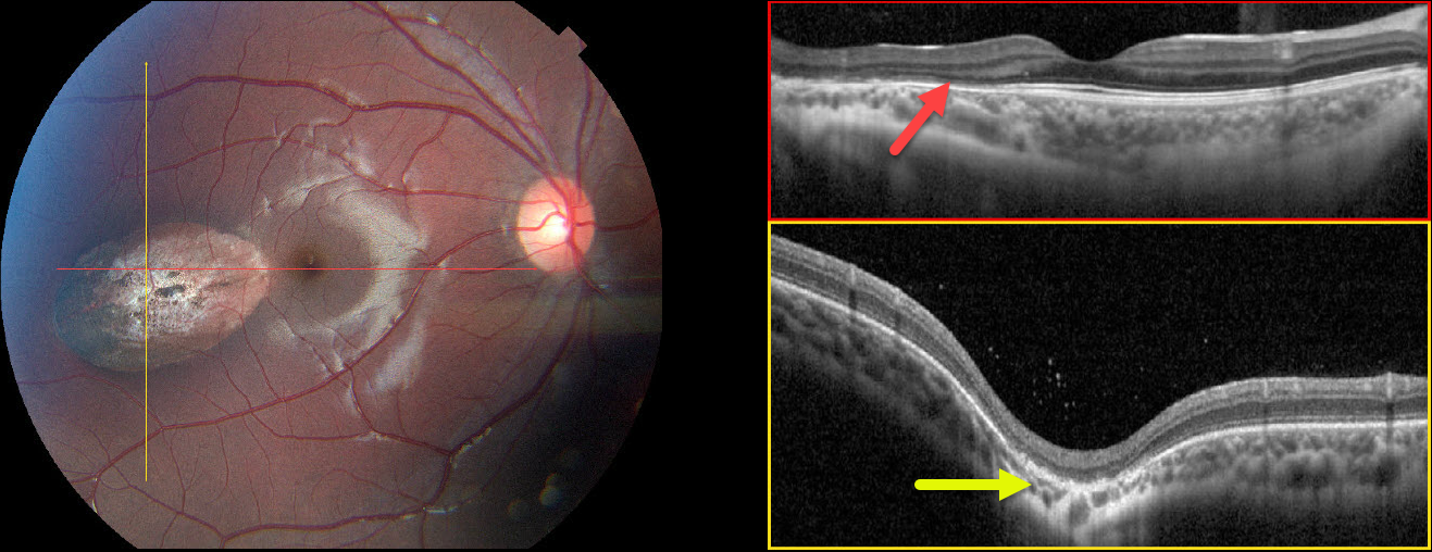
Color photography of her right eye shows an oval depigmented lesion in the temporal macula. Horizontal B-scan OCT shows outer retinal atrophy overlying the lesion. In the vertical B-scan, the lesion is colobomatous with outer retinal thinning. Some hyperreflective vitreous cells are noted. The left fundus was normal.
Learning Points:
Torpedo maculopathy was originally described by Gass as a solitary hypopigmented RPE nevus (Arch Ophthalmology 1992;110:1358-1359). These benign, unilateral lesions are tear-drop shaped and almost always located in the horizontal meridian in the temporal macula.
OCT scanning shows attenuation of the RPE and outer retinal layers. Vision is almost always normal, although there is a small risk for macular neovascularization. There are no known systemic associations.
Choroidal colobomas are most commonly due to failure of the optic vesicle and choroidal fissure to close during fetal development. Lesions like this may also be from other causes, including toxoplasmosis.
Although our patient’s lesion strongly resembles torpedo maculopathy, the coloboma would be highly atypical. Our best guess, given the vitreous cells, is that this lesion is due to toxoplasmosis.



